. 2018 Sep 30;5(11):1800776.
doi: 10.1002/advs.201800776. eCollection 2018 Nov.

Silicon-Enhanced Adipogenesis and Angiogenesis for Vascularized Adipose Tissue Engineering

Affiliations

Silicon-Enhanced Adipogenesis and Angiogenesis for Vascularized Adipose Tissue Engineering

Xiaoya Wang et al. Adv Sci (Weinh). .

Abstract

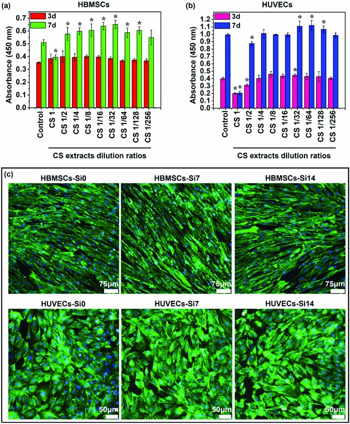
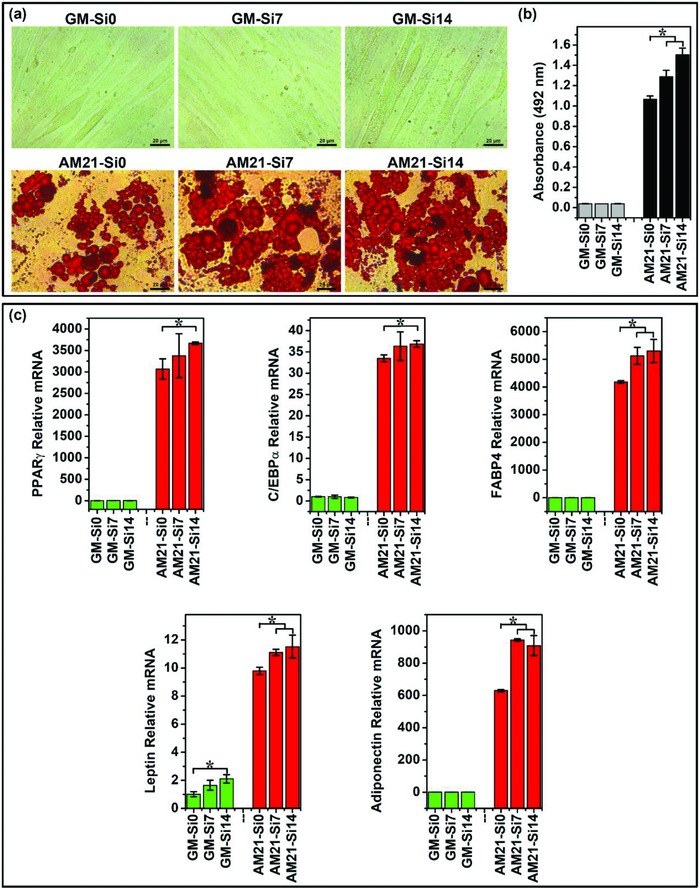
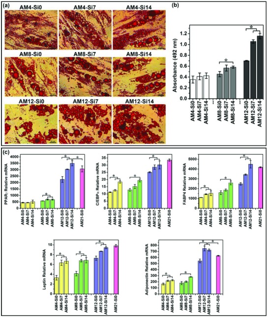
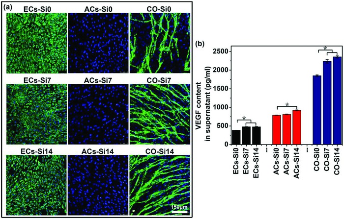
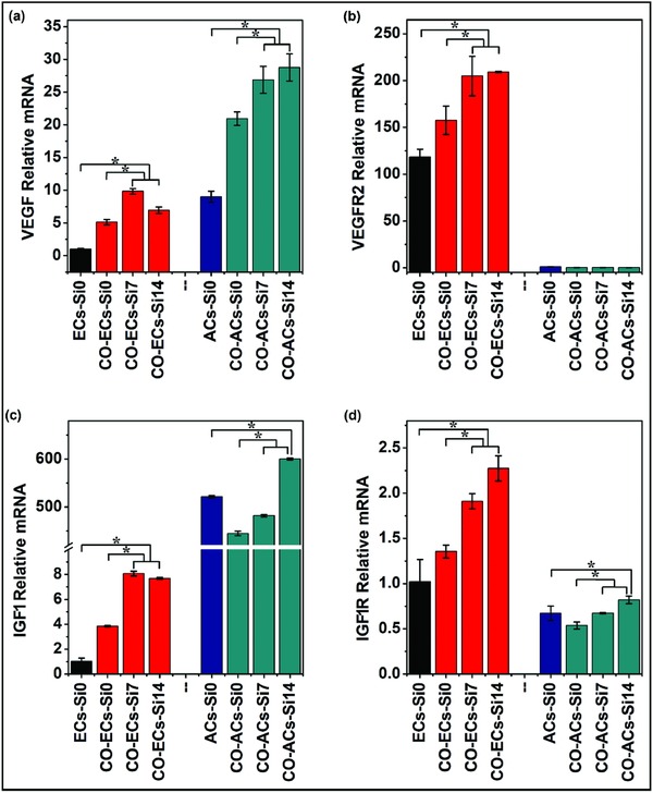
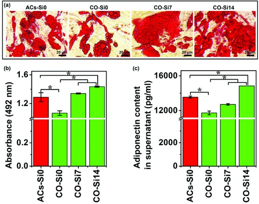
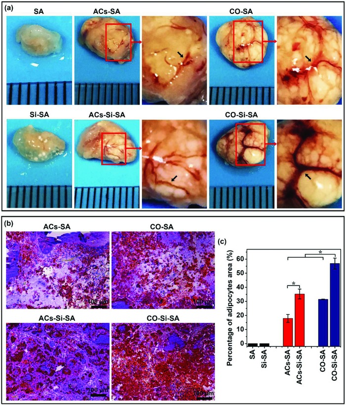
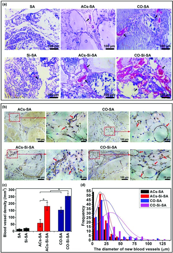
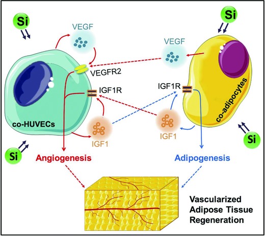
The enhancement of adipogenic differentiation of bone marrow mesenchymal stem cells (BMSCs) and sufficient vascularization remain great challenges for the successful reconstruction of engineered adipose tissue. Here, the bioactive effects of silicon (Si) ions on adipogenic differentiation of human BMSCs (HBMSCs) and the stimulation of vascularization during adipose tissue regeneration are reported. The results show that Si ions can enhance adipogenic differentiation of HBMSCs through the stimulation of the expression of adipogenic differentiation switches such as peroxisome proliferator-activated receptor γ and CCAAT/enhancer-binding protein α. Furthermore, Si ions can enhance both angiogenesis and adipogenesis, and inhibit dedifferentiation of cocultured adipocytes by regulating the interactions between HBMSC-derived adipocytes and human umbilical vein endothelial cells, in which the promotion of the expression of insulin-like growth factor 1 and vascular endothelial growth factor plays vital roles. The in vivo studies further demonstrate that the designed composite hydrogel with the ability to release bioactive Si ions clearly stimulates neovascularization and adipose tissue regeneration. The study suggests that Si ions released from biomaterials are important chemical cues for adipogenic differentiation and biomaterials with the ability to release Si ions can be designed for adipose tissue engineering.

Keywords: adipogenesis; adipogenic differentiation; adipose tissue engineering; angiogenesis; dedifferentiation.


Figures

Figure 1
Figure 1
Cell proliferation and morphology of HBMSCs and HUVECs cultured with different concentrations of Si ions. a,b) CCK‐8 assay indicated that Si ions in certain concentration range can stimulate the proliferation of HBMSCs and HUVECs (n = 6 for both the groups; *p < 0.05). c) Actin cytoskeleton staining assay showed that both concentrations of Si ions (7 and 14 µg mL−1) did not affect the morphology of HBMSCs and HUVECs. Scale bar, 75 µm for HBMSCs and 50 µm for HUVECs. The actin cytoskeletons were stained with FITC–Phalloidin (green), and nucleus was counterstained by using DAPI (blue).
Figure 2
Figure 2
Oil Red O staining and adipogenic gene expression of HBMSCs cultured in different mediums with or without Si ions. a) Oil Red O staining of cells showed that Si ions stimulated the increase of lipid accumulation. Lipid droplets were stained in red. Scale bar, 20 µm. b) Quantitative analysis of Oil Red O staining showed that Si ions significantly stimulated lipid accumulation (n = 3 for both the groups; *p < 0.05). c) Quantitative PCR analysis showed that Si ions enhanced the adipogenic marker gene of PPARγ, C/EBPα, FABP4, leptin, and adiponectin expression of HBMSCs (n = 3 for both the groups; *p < 0.05). GM‐Si0, GM‐Si7, GM‐Si14: HBMSCs cultured with growth medium containing different concentrations of Si ions (0, 7, and 14 µg mL−1) without differentiation inducer for 21 days, respectively; AM21‐Si0, AM21‐Si7, AM21‐Si14: HBMSCs cultured with adipogenic differentiation medium containing different concentrations of Si ions (0, 7, and 14 µg mL−1), respectively, for 21 days.
Figure 3
Figure 3
The effects of Si ion as a differentiation enhancer on the stimulation of adipogenic differentiation. a) Oil Red O staining of cells showed that Si ions stimulated the increase of lipid accumulation. Lipid droplets were stained in red. Scale bar, 20 µm. b) Quantitative analysis of Oil Red O staining showed that Si ions significantly stimulated lipid accumulation (n = 3 for both the groups; *p < 0.05). c) Quantitative PCR analysis showed that Si ions enhanced the adipogenic marker gene of PPARγ, C/EBPα, FABP4, leptin, and adiponectin expression of HBMSCs (n = 3 for both the groups; *p < 0.05). AM4‐Si7, AM4‐Si14: HBMSCs cultured with adipogenic differentiation medium alone for 4 days, and then further cultured up to 21 days in growth medium containing different concentrations of Si ions (0, 7, and 14 µg mL−1), respectively; AM8‐Si0, AM8‐Si7, AM8‐Si14: HBMSCs cultured with adipogenic differentiation medium alone for 8 days, and then further cultured up to 21 days in growth medium containing different concentrations of Si ions (0, 7, and 14 µg mL−1), respectively; AM12‐Si0, AM12‐Si7, AM12‐Si14: HBMSCs cultured with adipogenic differentiation medium alone for 12 days, and then further cultured up to 21 days in growth medium containing different concentrations of Si ions (0, 7, and 14 µg mL−1), respectively; AM21‐Si0: HBMSCs cultured in adipogenic differentiation medium without Si ions for 21 days.
Figure 4
Figure 4
Immunofluorescence staining of vWF and VEGF secretion of monocultured cells and cocultured cells treated with or without Si ions. a) vWF staining showed that clear capillary‐like networks were formed in the cocultured cells but not in the monocultured HUVECs and adipocytes. In addition, Si ions stimulated the network formation. vWF was stained in green and nuclei in blue with DAPI. Scale bar, 150 µm. b) Quantitative ELISA analysis showed that Si ions enhanced the VEGF secretion of monocultured cells and cocultured cells in culture medium (n = 3 for both the groups; *p < 0.05). ECs‐Si0, ECs‐Si7, ECs‐Si14: HUVECs (ECs) cultured with growth medium containing different concentrations of Si ions (0, 7, and 14 µg mL−1) for 3 days; ACs‐Si0, ACs‐Si7, ACs‐Si14: HBMSCs cultured in adipogenic differentiation medium with different concentrations of Si ions (0, 7, and 14 µg mL−1) for 15 days for the development of the adipocytes' (ACs) phenotype; CO‐Si0, CO‐Si7, CO‐Si14: HBMSCs first cultured in adipogenic differentiation medium with different concentrations of Si ions (0, 7, and 14 µg mL−1) for 12 days for adipogenic differentiation, and then cocultured with HUVECs containing different concentrations of Si ions (0, 7, and 14 µg mL−1) for 3 days.
Figure 5
Figure 5
Angiogenic and adipogenic gene expression of monocultured cells and cocultured cells treated with or without Si ions. HBMSCs were first cultured in adipogenic differentiation medium for 12 days for the development of the adipocyte phenotype, and then cocultured with HUVECs containing different concentrations of Si ions (0, 7, and 14 µg mL−1) for 3 days. a–d) Quantitative PCR analysis showed that Si ions stimulated the gene expression of VEGF, VEGFR2, IGF1, and IGF1R of cocultured HUVECs and cocultured adipocytes (n = 3 for both the groups; *p < 0.05). ECs‐Si0: monocultured HUVECs without Si ions; CO‐ECs‐Si0, CO‐ECs‐Si7, CO‐ECs‐Si14: cocultured HUVECs incubated with different concentrations of Si ions (0, 7, and 14 µg mL−1); ACs‐Si0: monocultured adipocytes without Si ions; CO‐ACs‐Si0, CO‐ACs‐Si7, CO‐ACs‐Si14: cocultured adipocytes incubated with different concentrations of Si ions (0, 7, and 14 µg mL−1).
Figure 6
Figure 6
Oil Red O staining and adiponectin secretion of monocultured cells and cocultured cells treated with or without Si ions. a) Oil Red O staining of cells showed that Si ions stimulated the increase of lipid accumulation in the coculture system and inhibited the dedifferentiation of cocultured adipocytes. Lipid droplets were stained in red. Scale bar, 20 µm. b) Quantitative analysis of Oil Red O staining (n = 3 for both the groups; *p < 0.05). c) Quantitative ELISA analysis showed that Si ions enhanced the adiponectin secretion of cocultured cells in culture medium (n = 3 for both the groups; *p < 0.05). ACs‐Si0: HBMSCs cultured in adipogenic differentiation medium without Si ions for 12 days for the development of the adipocytes' (ACs) phenotype; CO‐Si0, CO‐Si7, CO‐Si14: HBMSCs first cultured in adipogenic differentiation medium with different concentrations of Si ions (0, 7, and 14 µg mL−1) for 12 days for adipogenic differentiation, and then cocultured with HUVECs containing different concentrations of Si ions (0, 7, and 14 µg mL−1) for 3 days.
Figure 7
Figure 7
Tissue appearance and Oil Red O staining of engineered adipose tissue in a nude mice subcutaneous implant model by combining the monocultured cells or cocultured cells in calcium silicate/alginate composite hydrogel with the ability to release bioactive Si ions. a) Appearance of engineered adipose tissue showed that Si ions stimulated adipose tissue growth and vascular network (black arrows) formation. High magnification corresponded to boxed area (red) in the low magnification images. b) Oil Red O staining of implants showed that Si ions promoted adipose tissue regeneration in vivo. Lipid droplets were stained in red and nucleus were counterstained in blue with hematoxylin. Scale bar, 100 µm. c) Quantitative analysis of Oil Red O staining of the implants indicated that Si ions stimulated adipose tissue formation (n = 5 for both the groups; *p < 0.05). SA: hydrogels without cells; Si‐SA: Si ion–released hydrogels without cells; ACs‐SA: hydrogels with monocultured adipocytes; ACs‐Si‐SA: Si ion–released hydrogels with monocultured adipocytes; CO‐SA: hydrogels with cocultured adipocytes and HUVECs; CO‐Si‐SA: Si ion–released hydrogels with cocultured adipocytes and HUVECs.
Figure 8
Figure 8
H&E staining and CD31 immunohistochemical staining of engineered adipose tissue implants. a) H&E staining showed that Si ions stimulated neovascularization in vivo. Blood vessels (black arrows) was stained in red and nuclei in blue. Scale bar, 100 µm. b) CD31 immunohistochemical staining showed that Si ions stimulated numerous microvessels' formation. Blood vessels (red arrows) was stained in brown and nuclei in blue. High magnification corresponded to boxed area (red) in the low magnification images. Scale bar, 100 µm for low magnification groups, 20 µm for the high magnification groups. c) Quantitative analysis of blood vessel density of the implants (n = 5 for both the groups; *p < 0.05). d) Quantitative analysis of the vessel diameter indicated that Si ions stimulated the increase of vessel diameters (n = 112 for both the groups). SA: hydrogels without cells; Si‐SA: Si ion–released hydrogels without cells; ACs‐SA: hydrogels with monocultured adipocytes; ACs‐Si‐SA: Si ion–released hydrogels with monocultured adipocytes; CO‐SA: hydrogels with cocultured adipocytes and HUVECs; CO‐Si‐SA: Si ion–released hydrogels with cocultured adipocytes and HUVECs.
Figure 9
Figure 9
Illustration of the mechanisms of Si ions enhancing angiogenesis and adipogenesis for better vascularized adipose tissue regeneration.

Similar articles

Cited by

References

    1. a) Choi J. H., Gimble J. M., Lee K., Marra K. G., Rubin J. P., Yoo J. J., Vunjak‐Novakovic G., Kaplan D. L., Tissue Eng., Part B 2010, 16, 413; - PMC - PubMed
    2. b) Huber B., Volz A. C., Kluger P. J., Cell Tissue Res. 2015, 362, 269; - PubMed
    3. c) Volz A. C., Huber B., Kluger P. J., Differentiation 2016, 92, 52. - PubMed
    1. a) Prockop D. J., Science 1997, 276, 71; - PubMed
    2. b) Pittenger M. F., Mackay A. M., Beck S. C., Jaiswal R. K., Douglas R., Mosca J. D., Moorman M. A., Simonetti D. W., Craig S., Marshak D. R., Science 1999, 284, 143. - PubMed
    1. a) Ninomiya Y., Sugahara‐Yamashita Y., Nakachi Y., Tokuzawa Y., Okazaki Y., Nishiyama M., Biochem. Biophys. Res. Commun. 2010, 394, 303; - PubMed
    2. b) Gomillion C. T., Burg K. J., Biomaterials 2006, 27, 6052; - PubMed
    3. c) Masuda T., Furue M., Matsuda T., Tissue Eng. 2004, 10, 523; - PubMed
    4. d) Scott M. A., Nguyen V. T., Levi B., James A. W., Stem Cells Dev. 2011, 20, 1793. - PMC - PubMed
    1. a) Lu Z., Yuan Y., Gao J., Lu F., Cell Tissue Res. 2016, 364, 289; - PubMed
    2. b) Bellas E., Marra K. G., Kaplan D. L., Tissue Eng., Part C 2013, 19, 745. - PMC - PubMed
    1. Mauney J. R., Volloch V., Kaplan D. L., Biomaterials 2005, 26, 6167. - PubMed

LinkOut - more resources