

Distal respiratory tract viral infections in young children trigger a marked increase in alveolar mast cells
- PMID: 30480000
- PMCID: PMC6250563
- DOI: 10.1183/23120541.00038-2018
Distal respiratory tract viral infections in young children trigger a marked increase in alveolar mast cells
Abstract
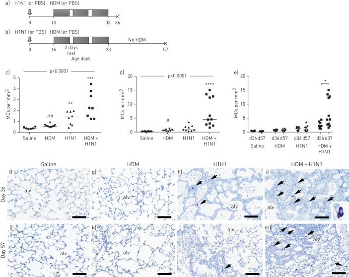
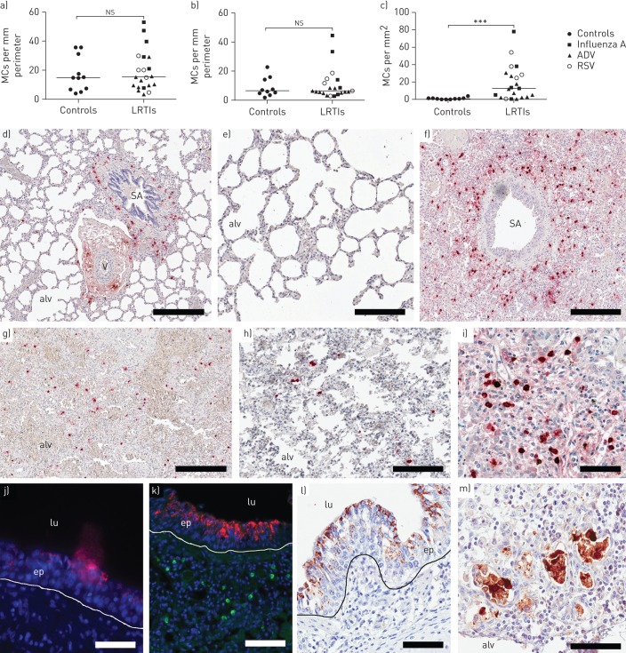
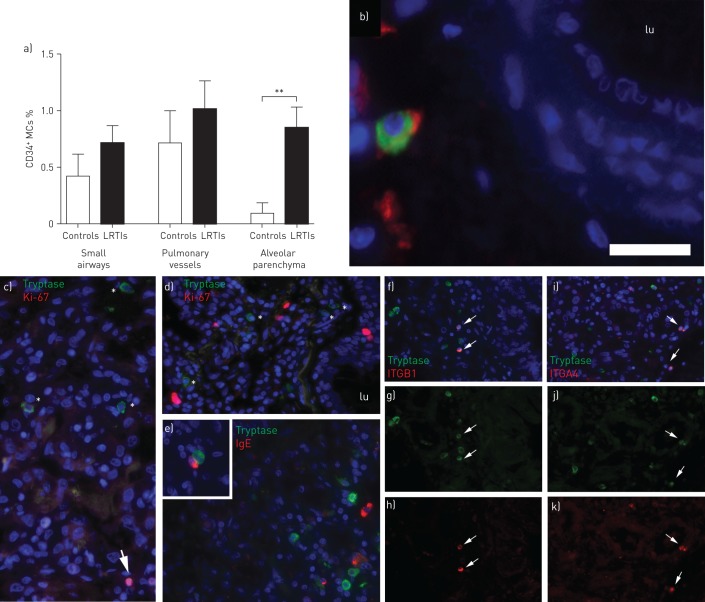
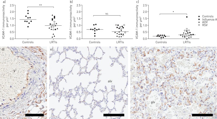
Viral infections predispose to the development of childhood asthma, a disease associated with increased lung mast cells (MCs). This study investigated whether viral lower respiratory tract infections (LRTIs) can already evoke a MC response during childhood. Lung tissue from young children who died following LRTIs were processed for immunohistochemical identification of MCs. Children who died from nonrespiratory causes served as controls. MCs were examined in relation to sensitisation in infant mice exposed to allergen during influenza A infection. Increased numbers of MCs were observed in the alveolar parenchyma of children infected with LRTIs (median (range) 12.5 (0-78) MCs per mm2) compared to controls (0.63 (0-4) MCs per mm2, p=0.0005). The alveolar MC expansion was associated with a higher proportion of CD34+ tryptase+ progenitors (controls: 0% (0-1%); LRTIs: 0.9% (0-3%) CD34+ MCs (p=0.01)) and an increased expression of the vascular cell adhesion molecule (VCAM)-1 (controls: 0.2 (0.07-0.3); LRTIs: 0.3 (0.02-2) VCAM-1 per mm2 (p=0.04)). Similarly, infant mice infected with H1N1 alone or together with house dust mite (HDM) developed an increase in alveolar MCs (saline: 0.4 (0.3-0.5); HDM: 0.6 (0.4-0.9); H1N1: 1.4 (0.4-2.0); HDM+H1N1: 2.2 (1.2-4.4) MCs per mm2 (p<0.0001)). Alveolar MCs continued to increase and remained significantly higher into adulthood when exposed to H1N1+HDM (day 36: 2.2 (1.2-4.4); day 57: 4.6 (1.6-15) MCs per mm2 (p=0.01)) but not when infected with H1N1 alone. Our data demonstrate that distal viral infections in young children evoke a rapid accumulation of alveolar MCs. Apart from revealing a novel immune response to distal infections, our data may have important implications for the link between viral infections during early childhood and subsequent asthma development.
Conflict of interest statement
Conflict of interest: C.K. Andersson has nothing to disclose. Conflict of interest: M. Shikhagaie has nothing to disclose. Conflict of interest: M. Mori has nothing to disclose. Conflict of interest: A. Al-Garawi has nothing to disclose. Conflict of interest: J.L. Reed has nothing to disclose. Conflict of interest: A.A. Humbles is an employee of and holds shares in MedImmune LLC. Conflict of interest: R. Welliver has nothing to disclose. Conflict of interest: T. Mauad has nothing to disclose. Conflict of interest: L. Bjermer has nothing to disclose. Conflict of interest: M. Jordana has nothing to disclose. Conflict of interest: J.S. Erjefält has nothing to disclose.
Figures
Similar articles
-
Influenza Infection in Mice Induces Accumulation of Lung Mast Cells through the Recruitment and Maturation of Mast Cell Progenitors.Front Immunol. 2017 Mar 22;8:310. doi: 10.3389/fimmu.2017.00310. eCollection 2017. Front Immunol. 2017. PMID: 28382037 Free PMC article.
-
Allergic airway inflammation induces migration of mast cell populations into the mouse airway.Cell Tissue Res. 2017 Aug;369(2):331-340. doi: 10.1007/s00441-017-2597-9. Epub 2017 Mar 25. Cell Tissue Res. 2017. PMID: 28343320
-
Allergin-1 on mast cells suppresses house dust mite-induced airway hyperresponsiveness in mice.Int Immunol. 2018 Aug 30;30(9):429-434. doi: 10.1093/intimm/dxy025. Int Immunol. 2018. PMID: 30169732
-
Uncontrolled asthmatics have increased FceRI+ and TGF-β-positive MCTC mast cells and collagen VI in the alveolar parenchyma.Clin Exp Allergy. 2018 Mar;48(3):266-277. doi: 10.1111/cea.13092. Epub 2018 Feb 15. Clin Exp Allergy. 2018. PMID: 29336501
-
Prevention of allergic disease in childhood: clinical and epidemiological aspects of primary and secondary allergy prevention.Pediatr Allergy Immunol. 2004 Jun;15 Suppl 16:4-5, 9-32. doi: 10.1111/j.1399-3038.2004.0148b.x. Pediatr Allergy Immunol. 2004. PMID: 15125698 Review.
Cited by
-
Defining mast cell differentiation and heterogeneity through single-cell transcriptomics analysis.J Allergy Clin Immunol. 2022 Oct;150(4):739-747. doi: 10.1016/j.jaci.2022.08.011. J Allergy Clin Immunol. 2022. PMID: 36205448 Free PMC article. Review.
-
Toll-like receptor activation induces airway obstruction and hyperresponsiveness in guinea pigs.Respir Res. 2024 Nov 29;25(1):421. doi: 10.1186/s12931-024-03050-3. Respir Res. 2024. PMID: 39614276 Free PMC article.
-
Expansion of Phenotypically Altered Dendritic Cell Populations in the Small Airways and Alveolar Parenchyma in Patients with Chronic Obstructive Pulmonary Disease.J Innate Immun. 2023;15(1):188-203. doi: 10.1159/000526080. Epub 2022 Aug 23. J Innate Immun. 2023. PMID: 35998572 Free PMC article.
-
Quantitative In-Depth Analysis of the Mouse Mast Cell Transcriptome Reveals Organ-Specific Mast Cell Heterogeneity.Cells. 2020 Jan 14;9(1):211. doi: 10.3390/cells9010211. Cells. 2020. PMID: 31947690 Free PMC article.
-
Respiratory Syncytial Virus Infection of Human Lung Fibroblasts Induces a Hyaluronan-Enriched Extracellular Matrix That Binds Mast Cells and Enhances Expression of Mast Cell Proteases.Front Immunol. 2020 Jan 28;10:3159. doi: 10.3389/fimmu.2019.03159. eCollection 2019. Front Immunol. 2020. PMID: 32047499 Free PMC article.
References
-
- Martinez FD. Viral infections and the development of asthma. Am J Respir Crit Care Med 1995; 151: 1644–1647. - PubMed
-
- Holt PG, Sly PD. Interaction between adaptive and innate immune pathways in the pathogenesis of atopic asthma: operation of a lung/bone marrow axis. Chest 2011; 139: 1165–1171. - PubMed
-
- Mackenzie KJ, Anderton SM, Schwarze J. Viral respiratory tract infections and asthma in early life: cause and effect? Clin Exp Allergy 2014; 44: 9–19. - PubMed
LinkOut - more resources
Full Text Sources
Miscellaneous