

Iatrogenic Paradoxical Stroke in a Patient With Catheter-Associated Thrombosis and Systemic-to-Pulmonary Venous Shunt
- PMID: 30480004
- PMCID: PMC6243407
- DOI: 10.1177/2324709618813175
Iatrogenic Paradoxical Stroke in a Patient With Catheter-Associated Thrombosis and Systemic-to-Pulmonary Venous Shunt
Abstract
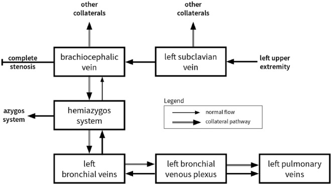
Paradoxical embolism occurs when thrombotic material traverses a right-to-left shunt. We describe the first case of paradoxical stroke resulting from manipulation of a disused chemotherapy port. Contrast studies revealed that the mechanism was systemic-to-pulmonary venous shunt, in which systemic veins drain into the left atrium via collaterals. Chronically thrombosed central venous catheters may result in venous stenosis and shunt formation, exposing patients to risks of paradoxical stroke, acute coronary syndrome, hypoxemia, and other complications. This case highlights the life-threatening complications that may result from neglect of an implantable central venous catheter. Preventative measures are to promptly recognize and treat catheter-related thrombosis and to remove unneeded catheters.
Keywords: central venous access device; paradoxical embolism; paradoxical stroke; right-to-left shunt; systemic-to-pulmonary venous shunt.
Conflict of interest statement
Declaration of Conflicting Interests: The author(s) declared no potential conflicts of interest with respect to the research, authorship, and/or publication of this article.
Figures


Similar articles
-
[Clinical characteristics of paradoxical brain embolism associated with isolated pulmonary arteriovenous fistula].Rinsho Shinkeigaku. 2002 Sep;42(9):849-54. Rinsho Shinkeigaku. 2002. PMID: 12710083 Japanese.
-
Right to left shunting through communications between the left superior intercostal vein tributaries and the left atrium: a potential cause of paradoxical embolism.Int J Cardiol. 2013 Sep 10;167(6):2867-74. doi: 10.1016/j.ijcard.2012.07.024. Epub 2012 Aug 9. Int J Cardiol. 2013. PMID: 22882965
-
Acute stroke, catheter related venous thrombosis, and paradoxical cerebral embolism: report of two cases.J Neuroimaging. 2013 Jan;23(1):111-4. doi: 10.1111/j.1552-6569.2010.00568.x. Epub 2011 Jan 31. J Neuroimaging. 2013. PMID: 21281383
-
Anomalous hepatic venous drainage into the left atrium: an unusual cause of hypoxemia.Respir Care. 2003 Jan;48(1):58-62. Respir Care. 2003. PMID: 12556263 Review.
-
Do central venous catheters have advantages over arteriovenous fistulas or grafts?J Nephrol. 2006 May-Jun;19(3):265-79. J Nephrol. 2006. PMID: 16874685 Review.
References
-
- Gilkeson RC, Nyberg EM, Sachs PB, Wiant AM, Zahka KG, Siwik ES. Systemic to pulmonary venous shunt: imaging findings and clinical presentations. J Thorac Imaging. 2008;23:170-177. - PubMed
-
- Murata K, Itoh H, Todo G, et al. Bronchial venous plexus and its communication with pulmonary circulation. Invest Radiol. 1986;21:24-30. - PubMed
LinkOut - more resources
Full Text Sources
