

Cervical skin denervation associates with alpha-synuclein aggregates in Parkinson disease
- PMID: 30480033
- PMCID: PMC6243385
- DOI: 10.1002/acn3.669
Cervical skin denervation associates with alpha-synuclein aggregates in Parkinson disease
Abstract
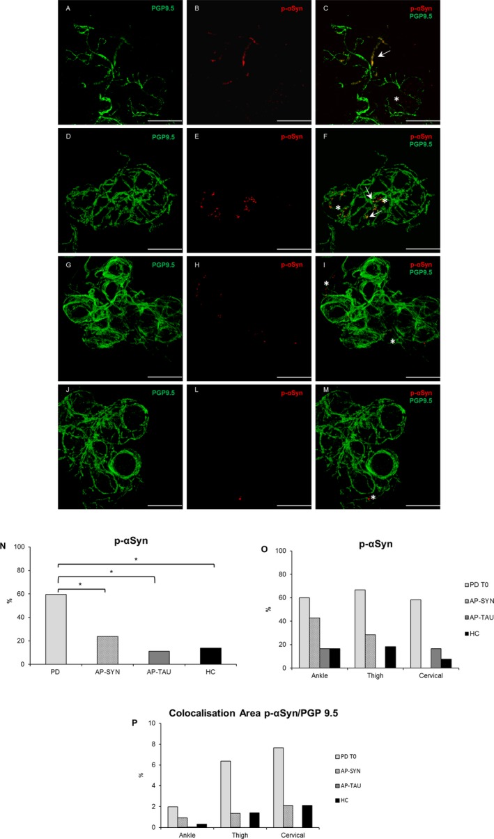
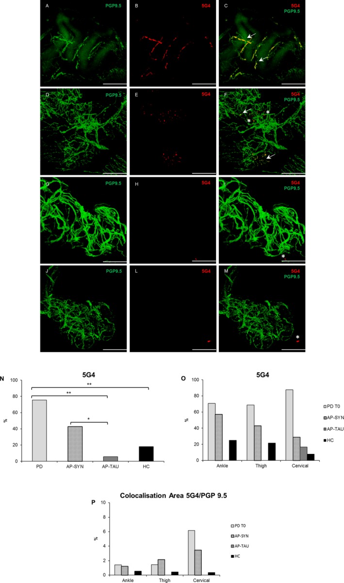
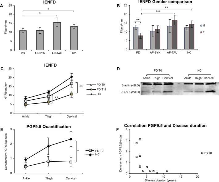
Objective: Autonomic nervous system is involved at the onset of Parkinson disease (PD), and alpha-synuclein (α-Syn) and its phosphorylated form (p-αSyn) have been detected in dermal autonomic nerve fibers of PD. We assessed disease specific conformation variant of α-Syn immunoreactivity in cutaneous nerves and characterized skin denervation patterns in PD and atypical parkinsonism (AP).
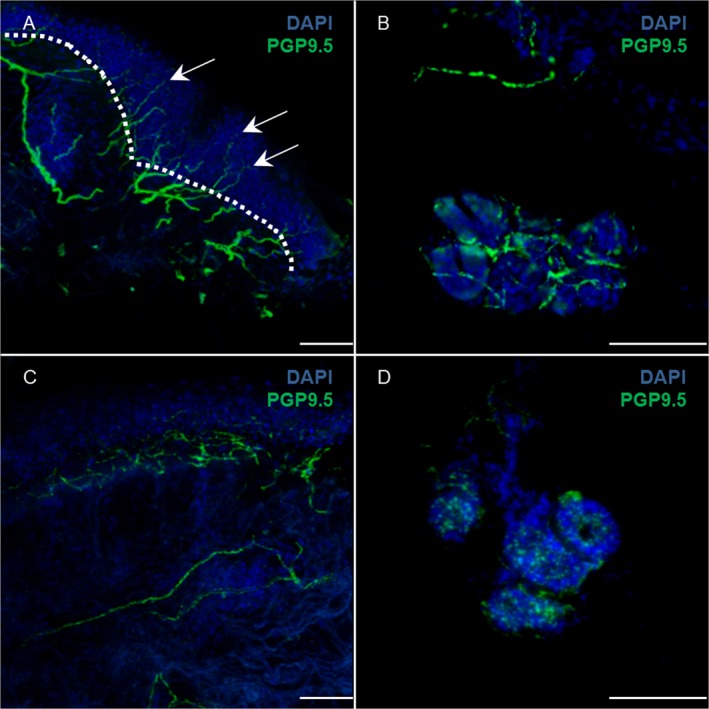
Methods: We enrolled 49 subjects, 19 with PD, 17 age-matched healthy controls, and 13 with AP. The manifestations of disease were rated on clinical scales. Skin biopsies from ankle, thigh, and neck were analyzed by immunofluorescence for p-αSyn, 5G4 as a conformation specific antibody to pathogenic α-Syn and PGP9.5 as axonal marker. Intraepidermal nerve fiber density was measured in all anatomical sites as marker of neurodegeneration. Thirteen of the 19 PD underwent a 1 year follow-up visit plus skin biopsies.
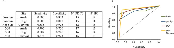
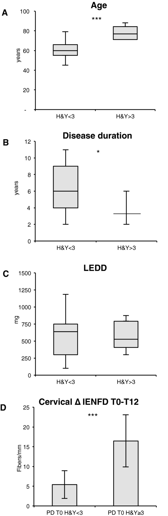
Results: PD subjects displayed more severe cervical skin denervation (P < 0.03), which correlated to disease duration and worsened between initial and follow-up examination (P < 0.001). p-αSyn and 5G4 were equally sensitive and specific for the diagnosis of PD (area under the ROC was 0.839 for p-αSyn and 0.886 for 5G4). PD and AP with possible alpha-synucleinopathies share the features of marked cervical denervation and the presence of 5G4. In contrast AP with possible tauopathies were normal.
Interpretation: Conformational specific forms of α-Syn are detectable in skin biopsy by immunofluorescence in PD, with a promising diagnostic efficiency similar to p-αSyn. Cervical cutaneous denervation correlates with disease duration and increases over time standing out as a potential biomarker of PD progression.
Figures
Similar articles
-
Alpha-synuclein oligomers and small nerve fiber pathology in skin are potential biomarkers of Parkinson's disease.NPJ Parkinsons Dis. 2021 Dec 20;7(1):119. doi: 10.1038/s41531-021-00262-y. NPJ Parkinsons Dis. 2021. PMID: 34930911 Free PMC article.
-
α-synuclein antibody 5G4 identifies idiopathic REM-sleep behavior disorder in abdominal skin biopsies.Parkinsonism Relat Disord. 2024 Mar;120:105956. doi: 10.1016/j.parkreldis.2023.105956. Epub 2023 Dec 9. Parkinsonism Relat Disord. 2024. PMID: 38217955
-
Phosphorylated α-Synuclein Deposits in Cutaneous Nerves of Early Parkinsonism.J Parkinsons Dis. 2022;12(8):2453-2468. doi: 10.3233/JPD-223421. J Parkinsons Dis. 2022. PMID: 36373295
-
Update on alpha-synuclein-based biomarker approaches in the skin, submandibular gland, gastrointestinal tract, and biofluids.Curr Opin Neurol. 2021 Aug 1;34(4):572-577. doi: 10.1097/WCO.0000000000000948. Curr Opin Neurol. 2021. PMID: 33967199 Review.
-
Skin nerve α-synuclein deposits in Parkinson's disease and other synucleinopathies: a review.Clin Auton Res. 2019 Dec;29(6):577-585. doi: 10.1007/s10286-018-0581-4. Epub 2018 Nov 30. Clin Auton Res. 2019. PMID: 30506233 Review.
Cited by
-
Proinflammatory profile in the skin of Parkinson's disease patients with and without pain.PLoS One. 2022 Oct 27;17(10):e0276564. doi: 10.1371/journal.pone.0276564. eCollection 2022. PLoS One. 2022. PMID: 36301901 Free PMC article.
-
Skin alpha-synuclein deposit patterns: A predictor of Parkinson's disease subtypes.EBioMedicine. 2022 Jun;80:104076. doi: 10.1016/j.ebiom.2022.104076. Epub 2022 May 26. EBioMedicine. 2022. PMID: 35644126 Free PMC article. Review.
-
Development of α-Synuclein Real-Time Quaking-Induced Conversion as a Diagnostic Method for α-Synucleinopathies.Front Aging Neurosci. 2021 Sep 28;13:703984. doi: 10.3389/fnagi.2021.703984. eCollection 2021. Front Aging Neurosci. 2021. PMID: 34650422 Free PMC article. Review.
-
Distribution of α-Synuclein Aggregation in the Peripheral Tissues.Neurochem Res. 2022 Dec;47(12):3627-3634. doi: 10.1007/s11064-022-03586-0. Epub 2022 Mar 28. Neurochem Res. 2022. PMID: 35348944 Review.
-
Alpha-synuclein oligomers and small nerve fiber pathology in skin are potential biomarkers of Parkinson's disease.NPJ Parkinsons Dis. 2021 Dec 20;7(1):119. doi: 10.1038/s41531-021-00262-y. NPJ Parkinsons Dis. 2021. PMID: 34930911 Free PMC article.
References
-
- Braak H, Rub U, Gai WP, Del Tredici K. Idiopathic Parkinson's disease: possible routes by which vulnerable neuronal types may be subject to neuroinvasion by an unknown pathogen. J Neural Transm (Vienna) 2003;110:517–536. - PubMed
-
- Kihara M, Kihara Y, Tukamoto T, et al. Assessment of sudomotor dysfunction in early Parkinson's disease. Eur Neurol 1993;33:363–365. - PubMed
-
- Swinn L, Schrag A, Viswanathan R, et al. Sweating dysfunction in Parkinson's disease. Mov Disord 2003;18:1459–1463. - PubMed
