. 2018 Sep 22;5(11):1408-1414.
doi: 10.1002/acn3.645. eCollection 2018 Nov.

Regulatory B cells in myasthenia gravis are differentially affected by therapies

Affiliations

Regulatory B cells in myasthenia gravis are differentially affected by therapies

Vuslat Yilmaz et al. Ann Clin Transl Neurol. .

Abstract

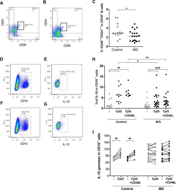
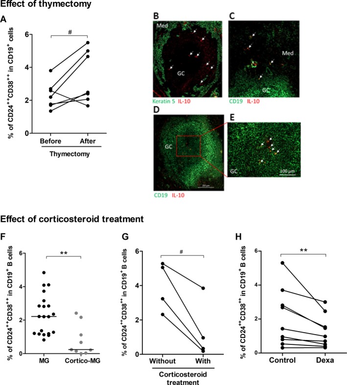
We analyzed the number and functionality of regulatory B (Breg) cells in well-defined myasthenia gravis patients. We first showed a decreased number of circulating CD19+ CD24++ CD38++ Breg cells and an altered functionality of Breg cells in untreated myasthenia gravis patients. Next, we demonstrated that the proportion of circulating Breg cells was restored in myasthenia gravis patients after thymectomy, probably as Breg cells could be sequestered in the myasthenia gravis thymus. In contrast, corticosteroid treatments did not restore and decreased even more the proportion of Breg cells in myasthenia gravis patients. These results clearly demonstrated that two distinct immunomodulatory therapies affect differentially Breg cells.


Figures

Figure 1
Figure 1
Altered number and function of peripheral Breg cells in MG. (A–C) PBMCs from untreated MG patients (n = 20) and healthy controls (n = 13) were labeled to identify Breg cells by flow cytometry. Representative FlowJo analyses of CD24++ CD38++ cells in CD19+ cells for a healthy donor (A) and an untreated MG patient (B). Percentage of CD24++ CD38++ cells in the CD19+ B cells (C). P‐values were assessed by the Mann–Whitney test (*P < 0.05). (D–I) PBMCs from untreated MG patients (n = 17–22) and healthy controls (n = 8–11) were cultivated 48 hours in resting condition or stimulated with CpG or CpG+CD40L. Representative FlowJo analyses for CD19+ IL‐10+ cells for a control donor in resting condition (D,E) or stimulated with CpG (F,G). PBLs were first gated for singlets on FSC‐H versus FSC‐A and for lymphocytes on SSC‐H vs FSC‐A. Next, CD19+ cells were analyzed in the lymphocyte gate (D,F) and the percentage and geomean for IL‐10‐positive cells in the CD19+ gate extracted (E,G). Global analyses of the percentage of IL‐10+ cells in the CD19 +  gate (H) and the geomean of fluorescence for IL‐10 in CD19+ cells (I) in control and MG cells. P‐values were assessed by the Wilcoxon paired test for comparison within each group (## P < 0.01, #### P < 0.001) and by the Mann–Whitney test for comparison between controls and MG patients (*P < 0.05, **P < 0.01).
Figure 2
Figure 2
Impact of classical MG therapies on the proportion of Breg cells. Effect of thymectomy: (A) PBMCs from seven MG patients were labeled before and 6–24 months after thymectomy to measure the percentage of CD24++ CD38++ in CD19+ B cells. P‐value was assessed by the paired t‐test (# P = 0.05). Only one patient had an inversed behavior with a decreased percentage of Breg cells after thymectomy. We had no reason to exclude the patient from the analysis but this patient also behave differentially regarding the % of regulatory T cells and Th17 cells compared to other patients. (B–E) Immunofluorescence staining of thymic sections from MG patients with an anti‐CD19 antibody for B cells or an anti‐keratin 5 for thymic medullary epithelial cells (in green), and anti‐IL10 (in red). Images were acquired with a Zeiss Axio Observer Z1 Inverted Microscope. Effect of corticosteroids: (F) PBMCs from MG patients (n = 20) and cortico‐treated MG patients (n = 9) were labeled to analyze the percentage of CD24++ CD38++ cells in the CD19+ B cells. P‐value was assessed by the Mann–Whitney test (**P = 0.002), (G) PBMCs from four MG patients followed up during in the course of their corticosteroid treatment were labeled to measure the percentage of CD24++ CD38++ in CD19+ B cells. P‐value was assessed by the paired t‐test (# P = 0.02). For three patients, the blood samples were initially without treatment and 1–24 months later while they were under corticosteroid treatment. For one patient, the blood sample was initially with corticosteroid treatment and 3 months later without treatment. (H) PBMCs from healthy controls were cultivated 48 hours with dexamethasone (Dexa) or without (control) and the percentages of CD24++ CD38++ cells in the CD19+ B cells were analyzed by flow cytometry. P‐value was assessed by the paired t‐test (**P = 0.008).

Similar articles

Cited by

References

    1. Berrih‐Aknin S, Le Panse R. Myasthenia gravis: a comprehensive review of immune dysregulation and etiological mechanisms. J Autoimmun 2014;52:90–100. - PubMed
    1. Wolfe GI, Kaminski HJ, Aban IB, et al. Randomized Trial of Thymectomy in Myasthenia Gravis. N Engl J Med 2016;375:511–522. - PMC - PubMed
    1. Mantegazza R, Bonanno S, Camera G, Antozzi C. Current and emerging therapies for the treatment of myasthenia gravis. Neuropsychiatr Dis Treat 2011;7:151–160. - PMC - PubMed
    1. Fillatreau S, Gray D, Anderton SM. Not always the bad guys: B cells as regulators of autoimmune pathology. Nat Rev Immunol 2008;8:391–397. - PubMed
    1. Rosser EC, Mauri C. Regulatory B cells: origin, phenotype, and function. Immunity 2015;42:607–612. - PubMed