Review
. 2018 Sep 27;5(11):1434-1455.
doi: 10.1002/acn3.658. eCollection 2018 Nov.

Clinical application of advanced MR methods in children: points to consider

Affiliations
Review

Clinical application of advanced MR methods in children: points to consider

Marko Wilke et al. Ann Clin Transl Neurol. .

Abstract

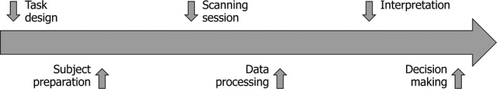
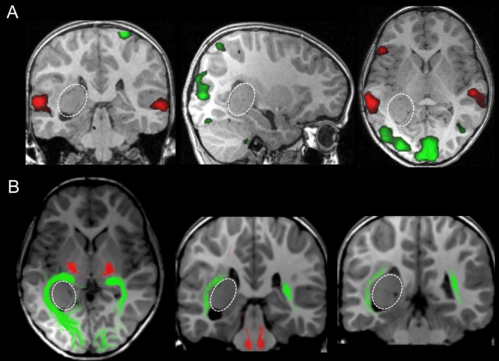
The application of both functional MRI and diffusion MR tractography prior to a neurosurgical operation is well established in adults, but less so in children, for several reasons. For this review, we have identified several aspects (task design, subject preparation, actual scanning session, data processing, interpretation of results, and decision-making) where pediatric peculiarities should be taken into account. Further, we not only systematically identify common issues, but also provide solutions, based on our experience as well as a review of the pertinent literature. The aim is to provide the clinician as well as the imaging scientist with information that helps to plan, conduct, and interpret such a clinically-indicated exam in a way that maximizes benefit for, and minimizes the burden on the individual child.


Figures

Figure 1
Figure 1
Illustration of the key aspects in the course of a clinically indicated functional MRI exam where pediatric peculiarities should be considered.
Figure 2
Figure 2
Illustration of the “triple use task” concept, with simple instructions to the child (“move your hands when you see the video, or listen to the story”). Here, assessing active condition 1 (C1, “video with moving hands” vs. “black screen and no moving hands”) induces activation in sensorimotor (S1/M1) and visual regions (V1/V5), while assessing active condition 2 (C2, “story with semantic violation” vs. “meaningless beep sounds”) induces activation in receptive (and partly, expressive) language regions (cf. Figure 3C and 6).
Figure 3
Figure 3
Illustration of clinically indicated fMRI exam for language in a 17‐year‐old boy with long‐standing epilepsy following a left‐frontal tumor removal in early childhood. Right‐hemispheric dominance was demonstrated for expressive (oval marker; vowel identification (A) and synonyms task (B)) and for receptive language (square marker; modified beep story (C), cf. Figure 2) and picture story task (D)). Also note consistent crossed cerebellar coactivation. Results are presented in neurological orientation, at P ≤ .001, uncorrected, with language activation in red and sensorimotor/visual activation in green.
Figure 4
Figure 4
Example of a miniature Lego MRI scanner model, allowing to prepare the child for the scanning session. Image courtesy of Julia Klebitz and with kind permission from “I love MRI” (http://www.amazings.eu/mri).
Figure 5
Figure 5
Illustration of clinically indicated fMRI/dMRI exam for motor system in a 6‐year‐old girl with a low‐grade glioma (oval marker). Depiction of functional activation in sensorimotor cortex (using active hand movement; (A) and functionally seeded depiction of the corresponding corticospinal tract (B), each being close to the lesion. Resection was planned accordingly and only a very minor facial palsy ensued. Results are presented in neurological orientation, at P ≤ 0.001, uncorrected, with sensorimotor activation in red. dMRI, diffusion MR tractography
Figure 6
Figure 6
Illustration of clinically indicated fMRI/dMRI exam for the visual system in a 6‐year‐old girl with a low‐grade glioma (oval marker). Depiction of functional activation in receptive language as well as primary and secondary visual brain regions (A), and the optic radiation (B). Demonstrating the bulged optic radiation lateral to the tumor allowed predicting a postoperative visual field defect, which in fact ensued following successful gross total tumor resection. Results are presented in neurological orientation, at p ≤ 0.001, uncorrected, with language activation in red and sensorimotor/visual activation in green (in A), and corticospinal tract in red and optic radiation in green (in B).

Similar articles

Cited by

References

    1. Schmidt MH, Marshall J, Downie J, Hadskis MR. Pediatric magnetic resonance research and the minimal‐risk standard. IRB 2011; 33, 1–6. - PubMed
    1. Barras CD, Asadi H, Baldeweg T, et al. Functional magnetic resonance imaging in clinical practice: state of the art and science. Aust Fam Physician 2016;45:798–803. - PubMed
    1. Bick AS, Mayer A, Levin N. From research to clinical practice: implementation of functional magnetic imaging and white matter tractography in the clinical environment. J Neurol Sci 2012;312:158–165. 10.1016/j.jns.2011.07.040. - DOI - PubMed
    1. Duncan JS, Winston GP, Koepp MJ, Ourselin S. Brain imaging in the assessment for epilepsy surgery. Lancet Neurol 2016;15:420–433. 10.1016/S1474-4422(15)00383-X. - DOI - PMC - PubMed
    1. Essayed WI, Zhang F, Unadkat P, et al. White matter tractography for neurosurgical planning: a topography‐based review of the current state of the art. Neuroimage Clin 2017;15:659–672. 10.1016/j.nicl.2017.06.011. - DOI - PMC - PubMed

LinkOut - more resources