. 2018 Apr:20:138-144.
doi: 10.1016/j.cobeha.2017.12.021. Epub 2018 Feb 3.

Acquisition, Maintenance, and Therapeutic Use of a Simple Motor Skill

Affiliations

Acquisition, Maintenance, and Therapeutic Use of a Simple Motor Skill

James J S Norton et al. Curr Opin Behav Sci. 2018 Apr.

Abstract

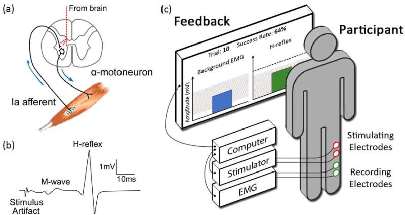
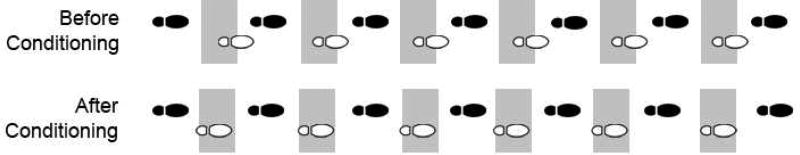
Operant conditioning of the spinal stretch reflex (SSR) or its electrical analog, the H-reflex, is a valuable experimental paradigm for studying the acquisition and maintenance of a simple motor skill. The CNS substrate of this skill consists of brain and spinal cord plasticity that operates as a hierarchy-the learning experience induces plasticity in the brain that guides and maintains plasticity in the spinal cord. This is apparent in the two components of the skill acquisition: task-dependent adaptation, reflecting brain plasticity; and long-term change, reflecting gradual development of spinal plasticity. The inferior olive, cerebellum, sensorimotor cortex, and corticospinal tract (CST) are essential components of this hierarchy. The neuronal and synaptic mechanisms of the spinal plasticity are under study. Because acquisition of this skill changes the spinal cord, it can affect other skills, such as locomotion. Thus, it enables investigation of how the highly plastic spinal cord supports the acquisition and maintenance of a broad repertoire of motor skills throughout life. These studies have resulted in the negotiated equilibrium model of spinal cord function, which reconciles the spinal cord's long-recognized reliability as the final common pathway for behaviors with its recently recognized ongoing plasticity. In accord with this model, appropriate H-reflex conditioning in a person with spasticity due to an incomplete spinal cord injury can trigger wider beneficial plasticity that markedly improves walking. H-reflex operant conditioning appears to provide a valuable new method for enhancing functional recovery in people with spinal cord injury and possibly other disorders as well.


Figures

Figure 1
Figure 1
(a) Main pathway of the spinal stretch reflex (SSR) and its electrical analog, the H-reflex. The pathway comprises the Ia afferent fiber from the muscle spindle, its synapse on the α-motoneuron, and the α-motoneuron itself. When the afferent is excited, it excites motoneurons innervating the muscle and its synergists. If it is excited by muscle stretch, the muscle response is the SSR; if it is excited by an electrical stimulus delivered to the nerve, the response is the H-reflex. The SSR and the H-reflex are typically measured by electromyography (EMG). While their pathway is entirely spinal, it is influenced by descending activity from the brain that affects the afferent synapse (presynaptically) and the motoneuron itself. Through this influence and the plasticity it induces in the spinal cord, the brain can gradually modify these spinal reflexes to increase rewards [11,46]. (b) Soleus EMG trace for a single trial, showing the stimulus artifact, the M-wave, and the H-reflex (modified from [13]). (c) Experimental setup for operant conditioning of the soleus H-reflex in human participants (based on experimental setups and figures in [9,12,18]). A pair of recording electrodes measures EMG activity from the soleus muscle; a pair of stimulating electrodes behind the knee elicits the H-reflex. The participant is asked to stand naturally and generate background EMG activity in the shaded range. After several seconds, a stimulus just above M-wave threshold elicits the M-wave and the H-reflex. (The threshold M-wave, or direct muscle response, results from excitation of a few large efferent fibers. Throughout the study, stimulus amplitude is continually adjusted to maintain the same M-wave size, thus ensuring that the effective stimulus strength does not change over the baseline, conditioning, and follow-up sessions.) If H-reflex size falls in the shaded range, the bar is green and the trial is a success; if it falls outside the range, the bar is red and the trial is not a success. The dashed horizontal line indicates the participant’s average H-reflex size from the control trials at the beginning of each experimental session. This illustration shows a successful down-conditioning trial; for up-conditioning the success range would be above a criterion.
Figure 2
Figure 2
Operant conditioning of the human soleus H-reflex. Data and method are from [9]. There were 3 stages (each with multiple sessions) to the protocol: baseline (6 sessions), conditioning (24 sessions), and follow-up (4 sessions). Baseline and conditioning sessions were usually scheduled three times a week for ten weeks. Follow-up sessions occurred 10–14 days, one month, two months, and three months after the final conditioning session. During each conditioning or follow-up session, the participant first completed a set of 20 control trials without instruction to change H-reflex size or being provided feedback on H-reflex size. S/he then completed three 75-trial blocks of conditioning trials in which s/he was asked to increase (or decrease) H-reflex size and was provided with feedback immediately after each trial indicating whether H-reflex size met the size criterion (Fig. 1c). (a) Total impact of the conditioning protocol on H-reflex size. Average H-reflex size for conditioning trials during each session (with standard error bars) for all successful up-conditioning participants (upward triangles) and down-conditioning participants (downward triangles) over the three stages of the study. The two components of skill acquisition are shown in b and c. (b) Task-dependent adaptation appears early and remains the same thereafter. It was separated from long-term change in H-reflex size by subtracting average H-reflex size for the control trials at the beginning of the session (i.e., (c)) from average H-reflex size for the conditioning trials of the session (i.e., (a)). Thus, it indicates the change in reflex size that the participant learns to produce immediately when s/he is asked to do so. This learning occurs over the first few conditioning sessions (i.e., over the first ~1000 conditioning trials). (c) Long-term change begins later and grows gradually thereafter. It is indicated by the average H-reflex size for the control trials of each session. The figure is simplified from [9], which has full details.
Figure 3
Figure 3
Brain and spinal cord structures (including spinal interneurons (IN)) involved in H-reflex operant conditioning and the connections between them. The pink ellipses indicate sites of plasticity in the spinal cord. The striped pink ellipses indicate putative sites of plasticity in the brain [26]. As described in the text, this multi-site plasticity is believed to function as a hierarchy in which the plasticity in the brain induces and maintains the plasticity in the spinal cord. (Figure modified and updated from [39] and [13].)
Figure 4
Figure 4
Down-conditioning of the soleus H-reflex improves step-cycle symmetry in a person with spasticity due to chronic incomplete spinal cord injury. The figure shows successive step cycles before and after down-conditioning. The open footprint shows the steps of the more impaired leg (i.e., the leg in which the soleus H-reflex was down-conditioned). The filled footprint shows the steps of the other leg. The shaded areas indicate where the steps of the impaired leg should occur (i.e., midway between the steps of the other leg). Before H-reflex down-conditioning, the steps of the impaired leg are delayed; thus, the subject is limping. After H-reflex down-conditioning, the steps occur on time; the limp is not evident. (Modified from [18].)

Similar articles

Cited by

References

    1. Compact Oxford English dictionary. second. Oxford University Press; 1993.
    1. Misiaszek JE. The H-reflex as a tool in neurophysiology: Its limitations and uses in understanding nervous system function. Muscle Nerve. 2003;28:144–160. - PubMed
    1. Pierrot-Deseilligny E, Burke D. The circuitry of the human spinal cord: spinal and corticospinal mechanisms of movement. Cambridge University Press; 2012. Outstanding comprehensive resource for scientists and clinicians studying spinal pathways and their plasticity. Includes authoritative descriptions of methodologies, spinal pathways, and the role of spinal pathways in movement.
    1. Tucker KJ, Tuncer M, Türker KS. A review of the H-reflex and M-wave in the human triceps surae. Hum Mov Sci. 2005;24:667–688. - PubMed
    1. Zehr PE. Considerations for use of the Hoffmann reflex in exercise studies. Eur J Appl Physiol. 2002;86:455–468. - PubMed

LinkOut - more resources