

Reactivation of latent HIV-1 in vitro using an ethanolic extract from Euphorbia umbellata (Euphorbiaceae) latex
- PMID: 30481211
- PMCID: PMC6258530
- DOI: 10.1371/journal.pone.0207664
Reactivation of latent HIV-1 in vitro using an ethanolic extract from Euphorbia umbellata (Euphorbiaceae) latex
Abstract
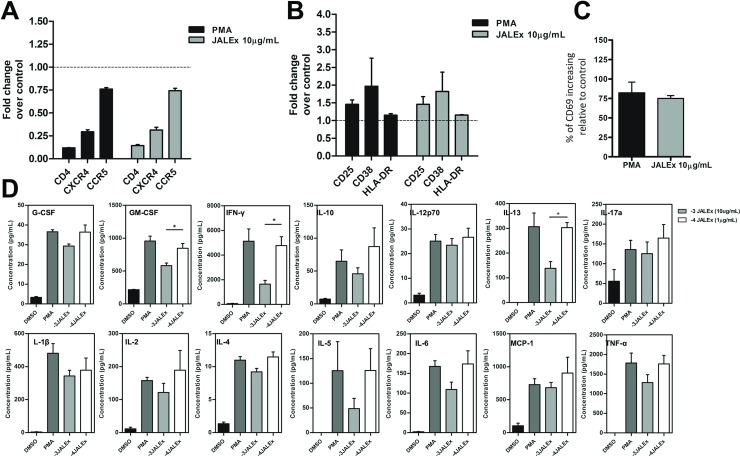
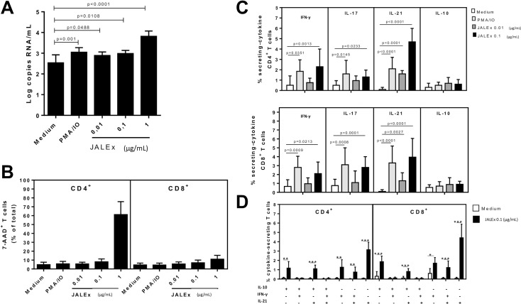
Euphorbia umbellata (E. umbellata) belongs to Euphorbiaceae family, popularly known as Janauba, and its latex contains a combination of phorbol esters with biological activities described to different cellular protein kinase C (PKC) isoforms. Here, we identified deoxi-phorbol esters present in E. umbellata latex alcoholic extract that are able to increase HIV transcription and reactivate virus from latency models. This activity is probably mediated by NF-kB activation followed by nuclear translocation and binding to the HIV LTR promoter. In addition, E. umbellata latex extract induced the production of pro inflammatory cytokines in vitro in human PBMC cultures. This latex extract also activates latent virus in human PBMCs isolated from HIV positive patients as well as latent SIV in non-human primate primary CD4+ T lymphocytes. Together, these results indicate that the phorbol esters present in E. umbellata latex are promising candidate compounds for future clinical trials for shock and kill therapies to promote HIV cure and eradication.
Conflict of interest statement
The authors have declared that no competing interests exist.
Figures
Similar articles
-
Phorbol Esters from the Latex of Euphorbia umbellata: Bioguided Isolation of Highly Potent HIV-1 Latency Interrupters in Virus Reservoir Cells.J Nat Prod. 2021 May 28;84(5):1666-1670. doi: 10.1021/acs.jnatprod.0c01092. Epub 2021 Apr 28. J Nat Prod. 2021. PMID: 33909429
-
Effects of diterpenes from latex of Euphorbia lactea and Euphorbia laurifolia on human immunodeficiency virus type 1 reactivation.Phytochemistry. 2010 Feb;71(2-3):243-8. doi: 10.1016/j.phytochem.2009.10.005. Epub 2009 Nov 10. Phytochemistry. 2010. PMID: 19897215
-
In vitro toxicity of latex, its terpenoidal fractions and isolated phorbol esters from Euphorbia umbellata (Pax) Bruyns on monocytic and melanoma cells.Fitoterapia. 2024 Jul;176:105987. doi: 10.1016/j.fitote.2024.105987. Epub 2024 May 3. Fitoterapia. 2024. PMID: 38703916
-
Bioactive Secondary Plant Metabolites from Euphorbia umbellata (PAX) BRUYNS (Euphorbiaceae).Chem Biodivers. 2022 Dec;19(12):e202200568. doi: 10.1002/cbdv.202200568. Epub 2022 Nov 15. Chem Biodivers. 2022. PMID: 36259393 Review.
-
The development of immune-modulating compounds to disrupt HIV latency.Cytokine Growth Factor Rev. 2012 Aug-Oct;23(4-5):159-72. doi: 10.1016/j.cytogfr.2012.05.003. Epub 2012 Jul 4. Cytokine Growth Factor Rev. 2012. PMID: 22766356 Review.
Cited by
-
Antiretroviral treatment, government policy and economy of HIV/AIDS in Brazil: is it time for HIV cure in the country?AIDS Res Ther. 2019 Aug 14;16(1):19. doi: 10.1186/s12981-019-0234-2. AIDS Res Ther. 2019. PMID: 31412889 Free PMC article. Review.
-
Euphorbia-Derived Natural Products with Potential for Use in Health Maintenance.Biomolecules. 2019 Aug 2;9(8):337. doi: 10.3390/biom9080337. Biomolecules. 2019. PMID: 31382529 Free PMC article. Review.
-
Bioactive Compounds from Euphorbia usambarica Pax. with HIV-1 Latency Reversal Activity.Pharmaceuticals (Basel). 2021 Jul 7;14(7):653. doi: 10.3390/ph14070653. Pharmaceuticals (Basel). 2021. PMID: 34358079 Free PMC article.
References
-
- Barré-Sinoussi F, Chermann JC, Rey F, Nugeyre MT, Chamaret S, Gruest J, et al. Isolation of a T-Lymphotropic Retrovirus from a Patient at Risk for Acquired Immune Deficiency Syndrome (AIDS). Science (80-). 1983;220: 868–871. - PubMed
-
- Gallo RC, Salahuddin SZ, Popovic M. Frequent Detection and Isolation of Cytopathic Retroviruses (HTLV-III) from Patients with AIDS and at Risk for AIDS Abstract. Science (80-). 1984;224: 3769–3772. - PubMed
-
- UNAIDS. 2017 Global Hiv Statistics. In: UNAIDS [Internet]. 2018 pp. 1–5. Available: http://www.unaids.org/sites/default/files/media_asset/UNAIDS_FactSheet_e...
-
- Levy JA. Virus-host interactions in HIV pathogenesis: directions for therapy. Adv Dent Res. 2011;23: 13–18. 10.1177/0022034511398874 - DOI - PMC - PubMed
-
- Coffin J, Swanstrom R. HIV pathogenesis: Dynamics and genetics of viral populations and infected cells. Cold Spring Harb Perspect Med. 2013;3 10.1101/cshperspect.a012526 - DOI - PMC - PubMed
Publication types
MeSH terms
Substances
LinkOut - more resources
Full Text Sources
Research Materials
