. 2019 Jan 25;47(2):806-823.
doi: 10.1093/nar/gky1180.

Role of the uS9/yS16 C-terminal tail in translation initiation and elongation in Saccharomyces cerevisiae

Affiliations

Role of the uS9/yS16 C-terminal tail in translation initiation and elongation in Saccharomyces cerevisiae

Supriya Jindal et al. Nucleic Acids Res. .

Abstract

The small ribosomal subunit protein uS9 (formerly called rpS16 in Saccharomyces cerevisiae), has a long protruding C-terminal tail (CTT) that extends towards the mRNA cleft of the ribosome. The last C-terminal residue of uS9 is an invariably conserved, positively charged Arg that is believed to enhance interaction of the negatively charged initiator tRNA with the ribosome when the tRNA is base-paired to the AUG codon in the P-site. In order to more fully characterize the role of the uS9 CTT in eukaryotic translation, we tested how truncations, extensions and substitutions within the CTT affect initiation and elongation processes in Saccharomyces cerevisiae. We found that uS9 C-terminal residues are critical for efficient recruitment of the eIF2•GTP•Met-tRNAiMet ternary complex to the ribosome and for its proper response to the presence of an AUG codon in the P-site during the scanning phase of initiation. These residues also regulate hydrolysis of the GTP in the eIF2•GTP•Met-tRNAiMet complex to GDP and Pi. In addition, our data show that uS9 CTT modulates elongation fidelity. Therefore, we propose that uS9 CTT is critical for proper control of the complex interplay of events surrounding accommodation of initiator and elongator tRNAs in the P- and A-sites of the ribosome.


Figures
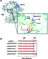
Figure 1.
Figure 1.
Structure and sequence analysis of uS9. (A) Location of ribosomal protein uS9 (formerly called S16 in yeast) in the head region of the small subunit (40S) of the eukaryotic (yeast) ribosome. The uS9 protein is depicted in navy blue and the 40S subunit is shown in grey. Inset: The CTT of uS9 is shown in navy blue with its last two amino acid residues, Tyrosine (Y) and Arginine (R), and the 40S ribosomal subunit shown in cyan. The Met-tRNAiMet is shown in green and mRNA is shown in red. The C-terminal Tyr and Arg residues in the uS9 CTT contact the anticodon stem loop of Met-tRNAiMet base paired with the AUG codon in the mRNA. PDB files 4V88 and 4KZZ were used for visualization using Swiss Pdbviewer. (B) C-terminal end sequences of the wild-type uS9 and uS9 C-terminal tail (CTT) mutants used in this study. Truncations, additions and substitutions introduced in uS9 are boxed.
Figure 2.
Figure 2.
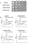
The uS9 C-terminal tail is essential for translation initiation in yeast cells. (A) Growth of wild-type and uS9 CTT mutant yeast strains (uS9/S16 R143Δ, uS9/S16 R143E, uS9/S16 R144). Cells were grown for 36 h on solid YEPD agar medium containing 2% glucose. (B) Translation initiation defects in uS9 mutants. Ribosome profiles of wild-type and mutant yeast strains. Whole cell extracts of the yeast strains were resolved by velocity sedimentation through 10–50% sucrose gradients. Fractions were collected while scanning at A254. The positions of different ribosomal species are indicated. Ratios of the area under the polyribosomal (P) and 80S (monosomal; M) peaks are shown (P:M) (mean ± standard error of the mean [SEM]).
Figure 3.
Figure 3.
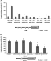
Changes in the length and charge of the uS9 CTT cause defects in translation reinitiation and resumption of scanning during GCN4 translation. (A) Translation re-initiation defects in uS9 mutants. Wild-type and mutant yeast strains were transformed with p180 GCN4-lacZ reporter construct and assayed for GCN4 re-initiation efficiency using 3-AT. p180 contains the wild-type GCN4 mRNA leader (all four uORFs). β-galactosidase activity (units) were measured under normal (without 3AT) and amino acid starved (+3AT) conditions. (B) GCN4-lacZ reporter activity. Wild-type and mutant yeast strains (uS9/S16-R143G, uS9/S16-R143Δ, uS9/S16-YRΔΔ, uS9/S16-R143E, uS9/S16-R144) were transformed with pM199 containing only uORF1, which is 140 nucleotides away from the GCN4 ORF. β-Galactosidase activity (units) was measured under normal conditions (-3AT) and is shown as the mean ±SEM from three biological replicates of three technical replicates each.
Figure 4.
Figure 4.
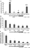
Changes in the length and charge of the uS9 CTT result in a leaky scanning phenotype as well as compromised AUG and UUG codon recognition. (A) GCN4-lacZ reporter activity in wild-type and mutant yeast strains (uS9/S16-R143G, uS9/S16-R143Δ, uS9/S16-YRΔΔ, uS9/S16-R143E, uS9/S16-R144) transformed with pM226 containing uORF1 extended into the GCN4 ORF. (B) Activity of HIS4-LacZ reporter constructs harboring AUG (B) or UUG (C) initiation codons following transformation into wild-type and mutant yeast strains. Mean β-galactosidase activity ± SEM determined from three biological replicates of three technical replicates each is shown.
Figure 5.
Figure 5.
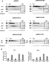
Association of eIF1 and eIF2α with 40S ribosomal subunits in wild-type and mutant (uS9/S16-R143G, uS9/S16-R143Δ, uS9/S16-YRΔΔ, uS9/S16-R143E, uS9/S16-R144) yeast strains. Extracts from isogenic wild-type and mutant strains were resolved by sucrose density gradient (10–30%) sedimentation. (A) Western blot analyses were performed using antibodies against eIF1, eIF2α and the ribosomal protein uS7/S5. Lanes marked ‘In’ for input contained a 7% portion of each gradient fraction. Analysis of eIF1 and eIF2α association was done using whole cell extract cross-linking with formaldehyde. (B) Association of eIF1 and eIF2 with 40S subunits was quantified and expressed as a percentage of 40S binding normalized against uS7.
Figure 6.
Figure 6.
The uS9/S16 C terminal region is important for eIF5-stimulated GTP hydrolysis. (A) Introduction of the TIF5-G31R allele reverses accumulation of eIF2α on uS9 mutant 40S ribosomal subunits. Association of initiation factor eIF2α with 40S subunits in uS9/S16-YRΔΔ, uS9/S16-R143E, uS9/S16-R144 (left panel) and uS9/S16-YRΔΔ <TIF5-G31R>, uS9/S16-R143E<TIF5-G31R>, uS9/S16-R144 <TIF5-G31R> (right panel) yeast strains. Western blot analysis of individual fractions with antibodies against eIF2 and uS7 is shown. ‘In’ for input - represents a 7% portion of each gradient fraction. (B) GTP hydrolysis by eIF2 with wild-type and mutant yeast 40S subunits. 40S•eIF1•eIF1A•mRNA (AUG) complexes were assembled in the presence of eIF5 and mixed with TC to initiate the GTP hydrolysis reaction.
Figure 7.
Figure 7.
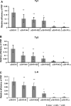
Reduced translation elongation fidelity in uS9/S16-R143G, uS9/S16-R143Δ, uS9/S16-YRΔΔ, uS9/S16-R143E, and uS9/S16-R144 strains. Wild-type (WT) and mutant yeast strains were transformed with (A) Ty1 (+1 frameshift reporter plasmid), (B) Ty3 (+1 frameshift reporter plasmid), and (C) L-A (–1 frameshift reporter plasmid). Dual luciferase assays were performed and programmed frameshifting (PRF) efficiencies were calculated as described in Materials and Methods. Mean efficiencies (relative to WT) determined from at least three independent experiments are plotted with bars representing standard errors. The statistical significance of differences in signals between mutant and WT strains is indicated.
Figure 8.
Figure 8.
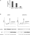
Antibiotic resistance and reduced eEF1A association of uS9 mutant yeast ribosomes. (A) Anisomycin resistance phenotypes of wild-type (WT) and uS9/S16-YRΔΔ mutant yeast strains. Overnight yeast cultures were diluted to OD 600 = 0.3, and 300 μl of the resulting suspensions were plated onto rich medium. A 0.5 cm diameter well was created sterilely in the center of the plate and filled with 20 μg anisomycin. Plates were incubated at 30°C for 3 days and the diameters of growth inhibition zones were monitored and plotted as bar graphs. (B) Cell extracts were resolved by velocity sedimentation on 7–50% sucrose gradients. Fractions were collected while scanning at A254 nm, resolved by SDS-PAGE and analyzed by Western blotting using antibodies against eEF1A and uS7. The positions of different ribosomal species are indicated.
Figure 9.
Figure 9.
Proposed model for uS9 C-terminal tail involvement in initiation and elongation processes in eukaryotes. (A) Initiation: Left—under wild-type conditions, proper positioning of the AUG start codon in the P-site is influenced by the correct location of the uS9 CTT and the charge of the last C-terminal positively charged Arg. The CTT triggers efficient eIF2-bound GTP → GDP + Pi hydrolysis, followed by optimal dissociation of eIF1 and eIF2 from the 48S complex. Right—reversal of the CTT C-terminal charge (red, –, uS9/S16-R143E mutant) and/or addition of an extra Arg (positive charge) (blue, ‡, uS9/S16-R144 mutant) results in inefficient GTP hydrolysis and compromised release of eIF1 and eIF2 from the complex. The severity of the effects of each mutation are reflected in the thickness of the dashed lines (thicker lines represent more severe defects while thinner lines represent less severe defects). (B) Elongation: Left - under wild-type conditions, the uS9 CTT mediates cooperation between the ribosomal P- and A-sites, promoting efficient eEF1A-mediated GTP hydrolysis and tRNA accommodation, followed by optimal dissociation of eEF1A. Right—deletions (uS9/S16-YRΔΔ mutant) and/or mutations in the CTT reduce cooperation between the P- and A-sites and result in more stringent tRNA selection/accommodation during elongation accompanied by enhanced eEF1A bound GTP-hydrolysis and dissociation of eEF1A from elongating 80S ribosomes.

Similar articles

Cited by

References

    1. Sonenberg N., Hinnebusch A.G.. Regulation of translation initiation in eukaryotes: mechanisms and biological targets. Cell. 2009; 136:731–745. - PMC - PubMed
    1. Ramakrishnan V. The ribosome emerges from a black box. Cell. 2014; 159:979–984. - PubMed
    1. Hinnebusch A.G. The scanning mechanism of eukaryotic translation initiation. Annu. Rev. Biochem. 2014; 83:779–812. - PubMed
    1. Jackson R.J., Hellen C.U., Pestova T.V.. The mechanism of eukaryotic translation initiation and principles of its regulation. Nat. Rev. Mol. Cell Biol. 2010; 11:113–127. - PMC - PubMed
    1. Dever T.E., Green R.. The elongation, termination, and recycling phases of translation in eukaryotes. Cold Spring Harb. Perspect. Biol. 2012; 4:a013706. - PMC - PubMed

Publication types

MeSH terms