. 2019 Apr;26(3):e12518.
doi: 10.1111/micc.12518. Epub 2019 Jan 15.

High salt diet impairs cerebral blood flow regulation via salt-induced angiotensin II suppression

Affiliations

High salt diet impairs cerebral blood flow regulation via salt-induced angiotensin II suppression

Linda A Allen et al. Microcirculation. 2019 Apr.

Abstract

Objectives: This study sought to determine whether salt-induced ANG II suppression contributes to impaired CBF autoregulation.

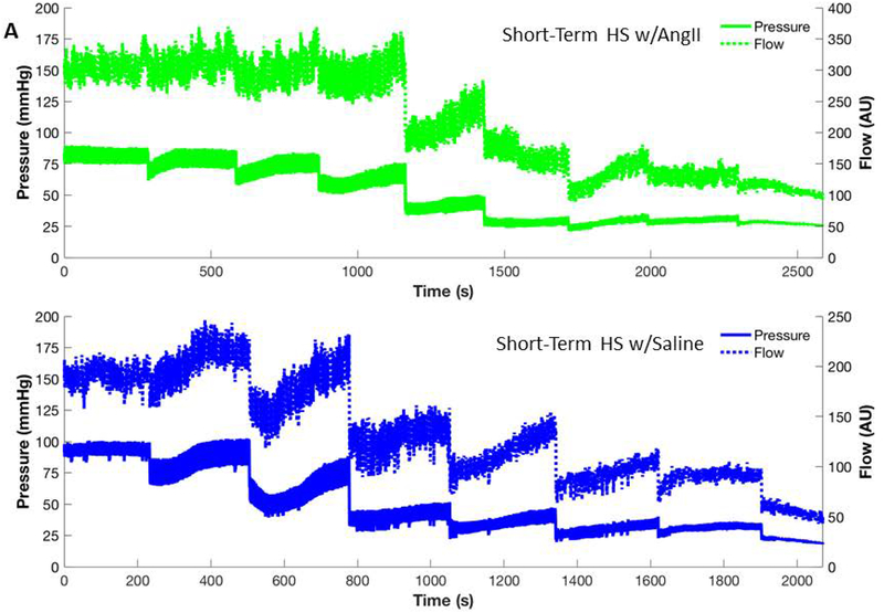
Methods: Cerebral autoregulation was evaluated with LDF during graded reductions of blood pressure. Autoregulatory responses in rats fed HS (4% NaCl) diet vs LS (0.4% NaCl) diet were analyzed using linear regression analysis, model-free analysis, and a mechanistic theoretical model of blood flow through cerebral arterioles.

Results: Autoregulation was intact in LS-fed animals as MAP was reduced via graded hemorrhage to approximately 50 mm Hg. Short-term (3 days) and chronic (4 weeks) HS diet impaired CBF autoregulation, as evidenced by progressive reductions of laser Doppler flux with arterial pressure reduction. Chronic low dose ANG II infusion (5 mg/kg/min, i.v.) restored CBF autoregulation between the pre-hemorrhage MAP and 50 mm Hg in rats fed short-term HS diet. Mechanistic-based model analysis showed a reduced myogenic response and reduced baseline VSM tone with short-term HS diet, which was restored by ANG II infusion.

Conclusions: Short-term and chronic HS diet lead to impaired autoregulation in the cerebral circulation, with salt-induced ANG II suppression as a major factor in the initiation of impaired CBF regulation.

Keywords: angiotensin II; autoregulation; cerebral blood flow; hemorrhage; salt.


Figures

Figure 1:
Figure 1:
Schematic representation of the mechanistic model of cerebral blood flow used to analyze the control and short-term high salt groups and the short-term high salt with saline infusion and short-term high salt with ANG II infusion groups. PA is the input arterial pressure, RAB is the resistance to flow of the cerebral arteries, CAB is the capacitance of the cerebral arteries, RATLB is the varying resistance to flow in the cerebral arterioles, CVB is the capacitance of the cerebral veins, RVB is the resistance to flow in the cerebral veins, and PVC is the pressure in the vena cava.
Figure 2:
Figure 2:
Representative blood pressure and cerebral blood flow response to serial blood withdrawals for control animals on a low salt diet (A) and a short-term (3 day) high salt diet (B). Each measurement is averaged using a moving window to see the trends in response of pressure and flow. In addition, during each of the blood withdrawals the pressure catheter was disconnected to allow blood to be withdrawn, so the periods during the actual blood withdrawal have been removed from the time course. Note that flow and pressure for the control animal (LS diet) does not change much over the first three blood withdrawals compared with changes observed in the short-term high salt animal. In addition, the control animal showed only ~ 25% drop in flow compared to ~ 75% drop in the short-term high salt animal over the course of the experiment. The shorter duration of the experimental period in the HS-fed rat vs. the LS-fed control reflects the reduced ability of the HS animal to compensate for the reduction in blood pressure following blood volume withdrawal.
Figure 3:
Figure 3:
Computational analysis of cerebral vascular autoregulation in rats fed low salt diet vs. short term HS diet. Panel A: Model free polynomial fit to each group. Panel B: Predicted average response of each group along with the predicted confidence intervals. Panel C: Optimized simulations of individual animal experiments showing that simulations of individual LS control rats predict a more distinct autoregulatory plateau than the simulations of the individual ST-HS rats, consistent with a rapid onset of impaired cerebral blood flow regulation following short term exposure to high salt diet (See text for details).
Figure 4:
Figure 4:
Representative blood pressure and cerebral blood flow response to serial blood withdrawals for animals on a short-term high salt diet with low dose ANG II infusion (A) and animals on a short-term high salt diet with saline infusion (B). Each measurement is averaged using a moving window to see the trends in response of pressure and flow. In addition, during each of the blood withdrawals the pressure catheter was disconnected to allow blood to be withdrawn so the periods during the actual blood withdrawal have been removed from the time course. Note that infusion of ANG II yields a response similar to the low salt control in Figure 2A, seemingly reversing the effects of a short-term high salt diet exhibited in saline-infused rats. The shorter duration of the experimental period in the HS animal receiving saline infusion vs. the HS-fed rat receiving low dose ANG II infusion reflects the improved ability of the ANG II-infused HS group to compensate for the reduction in blood pressure following blood volume withdrawal.
Figure 5:
Figure 5:
Computational analysis of cerebral vascular autoregulation in HS-fed rats receiving continuous i.v. infusion of a low dose of ANG II or continuous i.v. infusion of saline vehicle. Panel A: Model free polynomial fit to each group. Panel B: Predicted average response of each group along with the predicted confidence intervals. Panel C: Optimized simulations of individual animal experiments. The average response of the ST-HS + ANG II infusion group shows a distinct leftward shift in autoregulatory function when compared to the ST-HS + saline infusion group when analyzed with the mechanistic cerebral vasculature model (Figure 5B), while this distinction cannot be clearly seen in the model free analysis (Figure 5A) (See text for details).

References

    1. Amaral SL, Roman RJ, and Greene AS. Renin gene transfer restores angiogenesis and vascular endothelial growth factor expression in Dahl S rats. Hypertension 37: 386–390, 2001. - PubMed
    1. Barry DI, Strandgaard S, Graham DI, Braendstrup O, Svendsen UG, Vorstrup S, Hemmingsen R, and Bolwig TG. Cerebral blood flow in rats with renal and spontaneous hypertension: resetting of the lower limit of autoregulation. J Cereb Blood Flow Metab 2: 347–353, 1982. - PubMed
    1. Bohlen HG. Arteriolar closure mediated by hyperresponsiveness to norepinephrine in hypertensive rats. J Appl Physiol 236: H157–H164, 1979. - PubMed
    1. Cai H, Yao H, Ibayashi S, Zhao G, Kitazono T, Nagao T, and Fujishima M. Effects of long-acting angiotensin-converting enzyme inhibitor, imidapril, on the lower limit of cerebral blood flow autoregulation in hypertensive rats. Eur J Pharmacol 341: 73–77, 1998. - PubMed
    1. Campia U, Choucair WK, Bryant MB, Waclawiw MA, Cardillo C, and Panza JA. Reduced endothelium-dependent and -independent dilation of conductance arteries in African Americans. J Am Coll Cardiol 40: 754–760, 2002. - PubMed

Publication types

MeSH terms