. 2018 Nov 27;12(11):11041-11061.
doi: 10.1021/acsnano.8b05189. Epub 2018 Oct 16.

Breast Cancer Chemo-immunotherapy through Liposomal Delivery of an Immunogenic Cell Death Stimulus Plus Interference in the IDO-1 Pathway

Breast Cancer Chemo-immunotherapy through Liposomal Delivery of an Immunogenic Cell Death Stimulus Plus Interference in the IDO-1 Pathway

Jianqin Lu et al. ACS Nano. .

Retraction in

Abstract

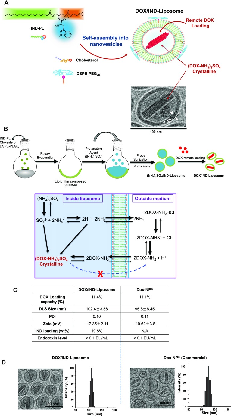
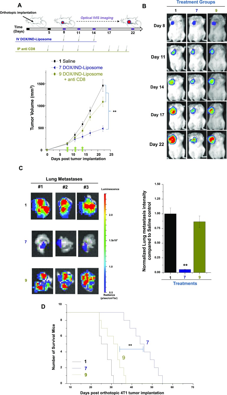
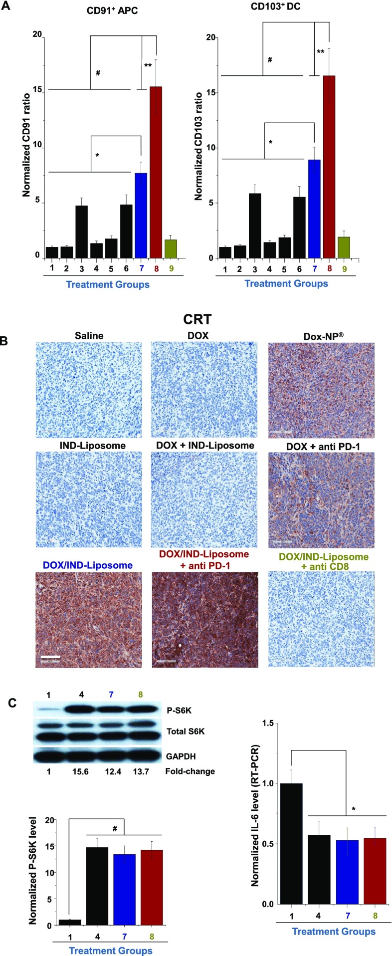
Immunotherapy provides the best approach to reduce the high mortality of metastatic breast cancer (BC). We demonstrate a chemo-immunotherapy approach, which utilizes a liposomal carrier to simultaneously trigger immunogenic cell death (ICD) as well as interfere in the regionally overexpressed immunosuppressive effect of indoleamine 2,3-dioxygenase (IDO-1) at the BC tumor site. The liposome was constructed by self-assembly of a phospholipid-conjugated prodrug, indoximod (IND), which inhibits the IDO-1 pathway, followed by the remote loading of the ICD-inducing chemo drug, doxorubicin (DOX). Intravenous injection of the encapsulated two-drug combination dramatically improved the pharmacokinetics and tumor drug concentrations of DOX and IND in an orthotopic 4T1 tumor model in syngeneic mice. Delivery of a threshold ICD stimulus resulted in the uptake of dying BC cells by dendritic cells, tumor antigen presentation and the activation/recruitment of naı̈ve T-cells. The subsequent activation of perforin- and IFN-γ releasing cytotoxic T-cells induced robust tumor cell killing at the primary as well as metastatic tumor sites. Immune phenotyping of the tumor tissues confirmed the recruitment of CD8+ cytotoxic T lymphocytes (CTLs), disappearance of Tregs, and an increase in CD8+/FOXP3+ T-cell ratios. Not only does the DOX/IND-Liposome provide a synergistic antitumor response that is superior to a DOX-only liposome, but it also demonstrated that the carrier could be effectively combined with PD-1 blocking antibodies to eradicate lung metastases. All considered, an innovative nano-enabled approach has been established to allow deliberate use of ICD to switch an immune deplete to an immune replete BC microenvironment, allowing further boosting of the response by coadministered IDO inhibitors or immune checkpoint blocking antibodies.

Keywords: breast cancer; chemo-immunotherapy; doxorubicin; dual-delivery liposome; immune checkpoint; immunogenic cell death; indoleamine 2,3-dioxygenase.


Conflict of interest statement

The authors declare the following competing financial interest(s): Andre E. Nel and Huan Meng are co-founders and equity holders in Westwood Biosciences Inc. The remaining authors declare no conflict of interest.

Figures

Figure 1
Figure 1
Schematic to explain BC immunotherapy by combined delivery of an immunogenic cell death stimulus plus an inhibitor of the IDO-1 pathway. Doxorubicin (DOX) delivery to the tumor site provides an effective stimulus for immunogenic cell death (ICD), which is characterized by calreticulin (CRT) expression (an “eat-me” signal for dendritic cell uptake) on the cancer cell surface. Subsequent release of adjuvant stimuli, HMGB-1 and ATP, by the dying cancer cells induce DC maturation and tumor antigen presentation to naïve T-cells. Recruitment of CD8+ cytotoxic T-lymphocytes (CTLs) triggers a full-fledged immune response, provided that the tumor infiltrating lymphocytes (TILs) can escape the immunosuppressive micromilieu at the BC tumor site. These immunosuppressive pathways include a contribution by FOXP-3+ regulatory T cells, autoregulatory effects of immune checkpoint receptors (e.g., PD-1) and the metabolic effects of the overexpressed IDO-1 immune surveillance pathway. The small molecule inhibitor, indoximod (IND), interferes in the IDO-1 pathway. We propose that simultaneous delivery of DOX and IND through the use of a nanocarrier can effectively combine the use of an ICD stimulus and interference in an immune surveillance pathway for the development of BC immunotherapy. The improved pharmacokinetics of drug delivery by the nanocarrier allows achievement of sufficiently high tumor drug levels to trigger an effective and sustained immune response for the reduction or elimination of the primary BC tumor and its metastases.
Figure 2
Figure 2
Use of a vaccination approach to identify chemo agents that induce ICD in a BC model. Published consensus guidelines were used to identify effective ICD introducing chemotherapy agents by a combination of in vitro 4T1 screening, followed by use of the dying tumor cells for a tumor vaccination procedure in syngeneic Balb/c mice. Multiparameter in vitro screening showed that doxorubicin (DOX) and paclitaxel (PTX), but not cisplatin (CIS), induced surface expression of CRT on 4T1 cells in a dose-dependent fashion, as well as quantifiable HMGB1 and ATP release (Figure S1B–D). (A) Animal vaccination, using 2 rounds of subcutaneous (SC) injection of dying 4T1 cells 7 days apart, followed by SC injection of live cells on the contralateral side. Successful growth inhibition at the challenge site is suggestive of immune interference. (B) Spaghetti plots showing growth inhibition of the tumors in animals vaccinated by dying tumor cells treated with DOX and PTX, but not CIS or PBS (n = 6). Evidence for the involvement of the innate and cognitive immune systems in the vaccination experiment appears in Figure S1F–H.
Figure 3
Figure 3
Synthesis of the dual-delivery DOX/IND-Liposome. (A) Schematic to show that the carrier is synthesized by self-assembly of an IND prodrug to form a liposome, which is subsequently loaded with DOX. The synthesis commences by conjugating IND to a single chain phospholipid [1-palmitoyl-2-hydroxy-sn-glycero-3-phosphocholine (PL)] to derive the IND-PL prodrug, as previously described by us. The three-step synthesis process is schematically depicted in Figure S3. The prodrug is mixed with cholesterol and DSPE-PEG2K to form a lipid film for the construction of liposomes. (B) Schematic to outline the liposome synthesis steps. Briefly, the lipid film comprised of IND-PL, cholesterol, and DSPE-PEG2K at a molar ratio of 70:25:5, was hydrated in a (NH4)2SO4 solution, followed by sonication and removal of free (NH4)2SO4. DOX was remotely loaded by using a proton gradient, as shown in the bottom panel. (NH4)2SO4 dissociates into protons, NH3 and SO42–. DOX is a weak basic molecule that is capable of diffusing across the IND-PL lipid bilayer into the liposome, where it is converted to a (DOX-NH3)2SO4 precipitate, incapable of back diffusion across the lipid bilayer. (C) Side-by-side comparison of DOX/IND-Liposome and Dox-NP for drug loading capacity, size, polydispersity, charge, and endotoxin levels (n = 3). (D) CryoEM pictures to show the morphological similarity between Dox-NP and IND-Liposome, including the presence of the drug precipitate.
Figure 4
Figure 4
Pharmacokinetics (PK) of drug delivery by the DOX/IND-Liposome compared to Dox-NP in an orthotopic tumor model. (A) Syngeneic orthotopic model was established injecting luciferase-transfected 4T1 cells into the 2nd mammary fat pad of Balb/c mice (left). This is followed by the development of a primary tumor mass that can be viewed by IVIS imaging after 2 weeks. Animal sacrifice and collecting the tumors and organs confirmed the treatment effect on primary tumor mass as well as the presence of metastatic nodules in the lung. After 4 weeks, it was possible to visualize the large primary tumor mass and extensive lung metastases. (B) Drug dose calculations for the animal studies (first n = 2, then n = 6, refer to the Methods section): maximum tolerated dose (MTD) calculations were carried out for the DOX formulations shown, using a NCI protocol. (C) IVIS imaging of DOX fluorescence at the 4T1 orthotopic tumor site (n = 3). Three animals in each group received free DOX, Dox-NP, and the DOX/IND-Liposome at 5 mg/kg DOX IV. The mice were sacrificed after 24 h for IVIS imaging. DOX fluorescence intensity was quantified by Living Image software. (D,E) PK and tissue drug distribution in 4T1 orthotopic tumor-bearing mice (n = 6), receiving IV injection of free DOX, Dox-NP, and DOX/IND-Liposome at a DOX equivalent dose of 5 mg/kg. Panel D depicts the WinNonlin software calculation of the plasma concentration, expressed as the % of the injected DOX dose at the indicated time points (left panel). The corresponding tumor and tissue drug concentrations, expressed as % injected DOX dose/g tissue, appears in the right panel. The equivalent data for IND appears in panel E. Results are expressed as mean ± SD; **p < 0.01 (ANOVA).
Figure 5
Figure 5
Treatment with the DOX/IND-Liposome impacts tumor growth and metastases in the orthotopic BC tumor model. (A) Outline of the experimental schedule (n = 9). The DOX/IND-Liposome was IV injected in animals in group 7 on days 8, 11, and 14 to deliver a DOX dose of 5 mg/kg and an IND dose of 8.7 mg/kg. Treatment was compared to IV injection of saline (#1), DOX (#2), Dox-NP (#3), (non-DOX-loaded) IND-PL-Liposome (#4), and DOX + IND-PL-Liposome (#5), using equivalent doses of DOX or IND. The inhibition of tumor growth by the DOX/IND-Liposome is significant compared to Dox-NP (*p < 0.05) and other controls (**p < 0.01, ANOVA). (B,C) Representative tumor images and average tumor weights after animal sacrifice on day 22. (D) Representative IVIS imaging and quantification of bioluminescence intensity of lung metastases (*p < 0.05; **p < 0.01, ANOVA). (E) Kaplan–Meier analysis to show that the DOX/IND-Liposome dramatically prolongs animal survival (n = 9, **p < 0.01, Log-rank Mantel-Cox test) in a separate experiment.
Figure 6
Figure 6
Anti-PD-1 coadministration with the DOX/IND-Liposome augments growth inhibition and eradication of lung metastases. Green arrows represent the treatment time points including DOX formulations and/or anti PD-1. (A) IHC staining showing pronounced PD-1 expression in the 4T1 BC tissue. (B) Tumor growth was assessed as in Figure 5, demonstrating that the addition of anti-PD-1 (injected IP at 100 μg/mouse on days 8, 11, and 14) exerted additional growth inhibitory effects (n = 9, ** p < 0.05 ANOVA). (C,D) Representative tumor images and tumor weights for the treatment groups. (E) Representative IVIS images and quantitative data to show the complete disappearance of lung metastases in animals receiving coadministration of anti-PD-1. (F) Kaplan–Meier analysis to show prolonged animal survival by anti-PD-1 administration (**p < 0.05, Log-rank Mantel-Cox test) in a separate experiment (n = 9).
Figure 7
Figure 7
Anti-CD8 monoclonal antibody interferes in the antitumor efficacy of the DOX/IND-Liposome. In order to demonstrate the critical role of cytotoxic CD8+ T-lymphocytes in antitumor immunity, anti-CD8 monoclonal was IP injected in treatment group 9, 3 days prior to the first drug administration and repeated every 2–3 days until the termination of the study. (A) Comparative tumor growth inhibition as described in Figure 5 (n = 9). (B) IVIS imaging data align with the growth inhibitory effects. (C) Representative ex vivo IVIS imaging, with quantification of luciferase expression, to show interference of anti-CD8 on lung metastatic spread. (D) Kaplan–Meier analysis to show that CD8 depletion dramatically reduces animal survival. Results are expressed as mean ± SD (n = 9, ** p < 0.01, ANOVA).
Figure 8
Figure 8
Immune phenotyping to demonstrate the effect of the DOX/IND-Liposome on initiating adaptive anti-BC immunity. Tumors were harvested from the different animal groups depicted in Table 1 to perform IHC staining and flow cytometry. (A) Multicolor flow cytometry analysis to show the impact on CD8/Treg ratios (n = 9). (B) IHC staining for CD8 expression in tumor tissue sections. (C) Flow cytometry analysis of T-cell IFN-γ+ expression in a CD45+CD3+CD8+IFN-γ+ gated cell population; granzyme B+ expression in a CD45+CD3+CD8+granzyme B+ gated cell population. (D) IHC staining for perforin expression. Additional IHC and flow data for the expression of FOXP-3, CC-3, IL12p70, and LC-3 are shown online (Figure S5C–E,G). The general treatment effect for the dual-delivery liposome (group 7) was to trigger effective cytotoxic killing at the tumor site, with significant boosting of the response by anti-PD-1 (group 8), and response decline during anti-CD8 administration (group 9).
Figure 9
Figure 9
Immune phenotyping to demonstrate the effect of the DOX/IND-Liposome on innate anti-BC immunity. The grouping order in this figure is identical to that of shown in Figure 8A. (A) Multicolor flow cytometry analysis to assess CD91 (CRT binding receptor) and CD103 expression on DCs (n = 9). (B) IHC staining to show CRT expression at the primary tumor site. Additional IHC and flow data for the expression of CD91 and CD80/CD86 are shown online (Figure S5F,H). (C) Western blotting to assess phosphorylation (activation) of P-S6 kinase in tumor tissues obtained from animal groups treated with the IND-Liposome (n = 3). The staining intensity of P-S6 kinase in relation to the amount of total kinase protein was graphically expressed by ImageJ software. The right panel shows RT-PCR analysis of IL-6 m-RNA levels at the tumor site. The increased phosphorylation of P-S6 kinase and reduced expression of IL-6 message RNA reflects interference of IND in the local immunosuppressive effects of IDO-1 that is overexpressed at the BC tumor site (Figure S2).
Figure 10
Figure 10
Encapsulated DOX delivery prevents toxicity of the heart, liver, and kidney. Blood was withdrawn on day 22 during performance of the efficacy studies in Figures 5–7 to assess the DOX-related toxicity in the (A) heart (troponin I and creatine kinase), (B) liver (ALT, AST), and kidney (creatinine). Encapsulated DOX delivery by Dox-NP and the DOX/IND-Liposome reduced the free drug toxicity. Results are expressed as mean ± SD (n = 9, *p < 0.05; **p < 0.01, ANOVA).

Similar articles

Cited by

References

    1. Edwards B. K.; Ward E.; Kohler B. A.; Eheman C.; Zauber A. G.; Anderson R. N.; Jemal A.; Schymura M. J.; Lansdorp-Vogelaar I.; Seeff L. C.; van Ballegooijen M. C.; Goede S. L.; Ries L. A. G. Annual Report to the Nation on the Status of Cancer, 1975–2006, Featuring Colorectal Cancer Trends and Impact of Interventions (risk factors, screening, and treatment) to Reduce Future Rates. Cancer 2010, 116, 544–573. 10.1002/cncr.24760. - DOI - PMC - PubMed
    1. Rashid O. M.; Takabe K. Does Removal of the Primary Tumor in Metastatic Breast Cancer Improve Survival?. J. Womens Health 2014, 23, 184–188. 10.1089/jwh.2013.4517. - DOI - PMC - PubMed
    1. Pardoll D. M. The Blockade of Immune Checkpoints in Cancer Immunotherapy. Nat. Rev. Cancer 2012, 12, 252–264. 10.1038/nrc3239. - DOI - PMC - PubMed
    1. Sharma P.; Allison J. P. The Future of Immune Checkpoint Therapy. Science 2015, 348, 56–61. 10.1126/science.aaa8172. - DOI - PubMed
    1. Sharma P.; Wagner K.; Wolchok J. D.; Allison J. P. Novel Cancer Immunotherapy Agents with Survival Benefit: Recent Successes and Next steps. Nat. Rev. Cancer 2011, 11, 805–812. 10.1038/nrc3153. - DOI - PMC - PubMed

Publication types

MeSH terms