

SLC25A22 Promotes Proliferation and Metastasis of Osteosarcoma Cells via the PTEN Signaling Pathway
- PMID: 30482097
- PMCID: PMC6259056
- DOI: 10.1177/1533033818811143
SLC25A22 Promotes Proliferation and Metastasis of Osteosarcoma Cells via the PTEN Signaling Pathway
Abstract
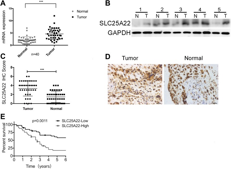
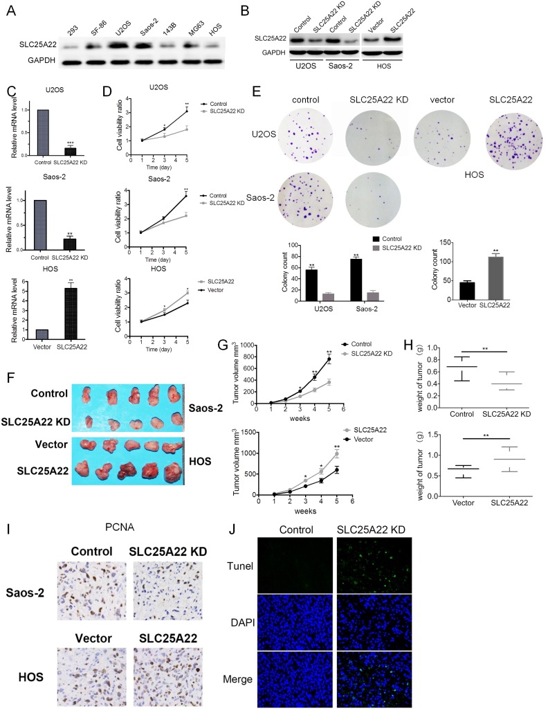
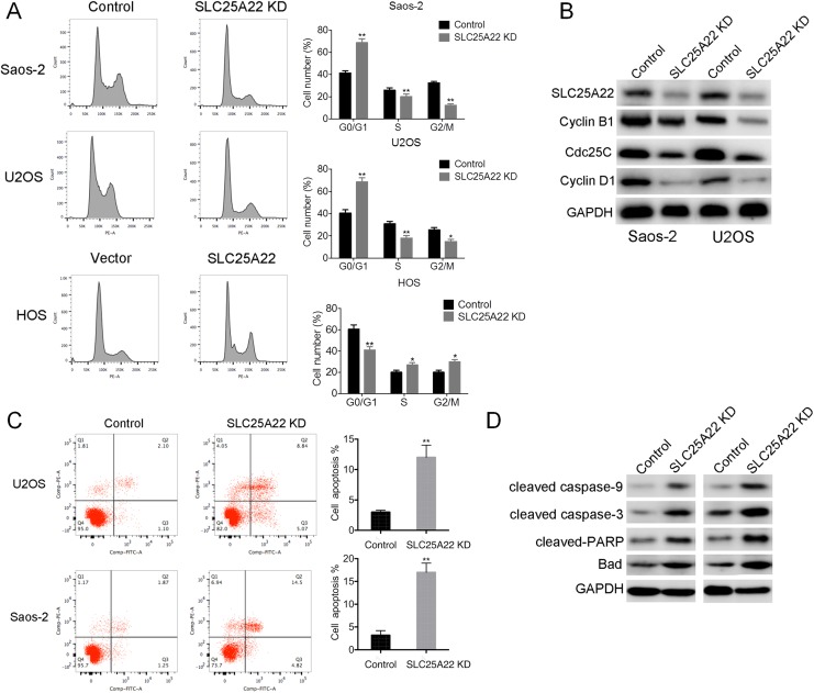
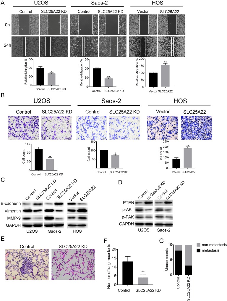
Osteosarcoma is a highly malignant bone tumor. However, due to the high complexity of the occurrence and metastasis of osteosarcoma, the exact mechanism promoting its development and progression remains to be elucidated. This study highlights the causal link between solute carrier family 25 member 22 (SLC25A22) and the development, progression, and metastasis of osteosarcoma. SLC25A22 is upregulated in human osteosarcoma and predicts a poor prognosis. The upregulation of SLC25A22 in osteosarcoma tissues was significantly associated with cell proliferation, invasion, and metastasis. Studies of functional gain (overexpression) and loss (knockdown) showed that SLC25A22 significantly increases the ability of osteosarcoma cells to proliferate, as well as invade and metastasize in vitro. At the same time, the expression of SLC25A22 promoted the progression of the cellcycle of osteosarcoma cell lines and inhibited the apoptosis of osteosarcoma cells. Analysis using a mouse xenograft model showed that xenografts of SLC25A22 stable overexpressing osteosarcoma cells had a significant increase in tumor volume and weight compared to the control group. Lung metastasis models in mice showed that expression of SLC25A22 promoted lung metastasis of osteosarcoma in vivo. Furthermore, SLC25A22 inhibited phosphatase and tensin homolog expression and increased phosphorylation of protein kinase b (Akt) and Focal Adhesion Kinase (FAK) in the phosphatase and tensin homolog signaling pathway. In summary, SLC25A22 is highly expressed in osteosarcoma, promoting osteosarcoma cell proliferation and invasion by inhibiting the phosphatase and tensin homolog signaling pathway.
Keywords: PTEN signaling pathway; SLC25A22; metastasis; osteosarcoma; proliferation.
Conflict of interest statement
Figures
Similar articles
-
MicroRNA-1908 is upregulated in human osteosarcoma and regulates cell proliferation and migration by repressing PTEN expression.Oncol Rep. 2015 Nov;34(5):2706-14. doi: 10.3892/or.2015.4242. Epub 2015 Sep 1. Oncol Rep. 2015. PMID: 26328886
-
SLC25A22 Promotes Proliferation and Survival of Colorectal Cancer Cells With KRAS Mutations and Xenograft Tumor Progression in Mice via Intracellular Synthesis of Aspartate.Gastroenterology. 2016 Nov;151(5):945-960.e6. doi: 10.1053/j.gastro.2016.07.011. Epub 2016 Jul 21. Gastroenterology. 2016. PMID: 27451147
-
MicroRNA-92a promotes epithelial-mesenchymal transition through activation of PTEN/PI3K/AKT signaling pathway in non-small cell lung cancer metastasis.Int J Oncol. 2017 Jul;51(1):235-244. doi: 10.3892/ijo.2017.3999. Epub 2017 May 16. Int J Oncol. 2017. PMID: 28534966
-
PTEN in osteosarcoma: Recent advances and the therapeutic potential.Biochim Biophys Acta Rev Cancer. 2020 Dec;1874(2):188405. doi: 10.1016/j.bbcan.2020.188405. Epub 2020 Aug 19. Biochim Biophys Acta Rev Cancer. 2020. PMID: 32827577 Review.
-
Bone Microenvironment and Osteosarcoma Metastasis.Int J Mol Sci. 2020 Sep 23;21(19):6985. doi: 10.3390/ijms21196985. Int J Mol Sci. 2020. PMID: 32977425 Free PMC article. Review.
Cited by
-
Role of Mitochondrial Transporters on Metabolic Rewiring of Pancreatic Adenocarcinoma: A Comprehensive Review.Cancers (Basel). 2023 Jan 8;15(2):411. doi: 10.3390/cancers15020411. Cancers (Basel). 2023. PMID: 36672360 Free PMC article. Review.
-
Glutamine-Derived Aspartate Biosynthesis in Cancer Cells: Role of Mitochondrial Transporters and New Therapeutic Perspectives.Cancers (Basel). 2022 Jan 4;14(1):245. doi: 10.3390/cancers14010245. Cancers (Basel). 2022. PMID: 35008407 Free PMC article. Review.
-
Identification of a Solute Carrier Family-Based Signature for Predicting Overall Survival in Osteosarcoma.Front Genet. 2022 Apr 19;13:849789. doi: 10.3389/fgene.2022.849789. eCollection 2022. Front Genet. 2022. PMID: 35518353 Free PMC article.
-
Identification of Some Glutamic Acid Derivatives with Biological Potential by Computational Methods.Molecules. 2023 May 16;28(10):4123. doi: 10.3390/molecules28104123. Molecules. 2023. PMID: 37241864 Free PMC article.
-
Osteosarcoma and Metastasis.Front Oncol. 2021 Dec 10;11:780264. doi: 10.3389/fonc.2021.780264. eCollection 2021. Front Oncol. 2021. PMID: 34956899 Free PMC article. Review.
References
-
- Pruksakorn D, Phanphaisarn A, Pongnikorn D, et al. AgeStandardized incidence rates and survival of osteosarcoma in northern Thailand. Asian Pac J Cancer Prev. 2016;17(7):3455–3458. - PubMed
-
- Marina N, Gebhardt M, Teot L, Gorlick R. Biology and therapeutic advances for pediatric osteosarcoma. Oncologist. 2004;9(4):422–441. - PubMed
-
- Chou AJ, Geller DS, Gorlick R. Therapy for osteosarcoma: where do we go from here? Paediatr Drugs. 2008;10(5):315–327. - PubMed
MeSH terms
Substances
LinkOut - more resources
Full Text Sources
Molecular Biology Databases
Research Materials
Miscellaneous
