. 2018 Nov 27;18(1):372.
doi: 10.1186/s12887-018-1346-x.

Necrotizing enterocolitis leads to disruption of tight junctions and increase in gut permeability in a mouse model

Affiliations

Necrotizing enterocolitis leads to disruption of tight junctions and increase in gut permeability in a mouse model

Srikanth Ravisankar et al. BMC Pediatr. .

Abstract

Background: Necrotizing enterocolitis (NEC) is a leading cause of death in preterm infants. Neonates weighing <1500 grams are at the highest risk for acquiring NEC, with a prevalence of nearly 7-10%, mortality up to 30%, and several long-term complications among survivors. Despite advancements in neonatal medicine, this disease remains a challenge to treat. The aim of this study is to investigate the effect of NEC on gut epithelial tight junctions and its barrier function using a NEC mouse model.

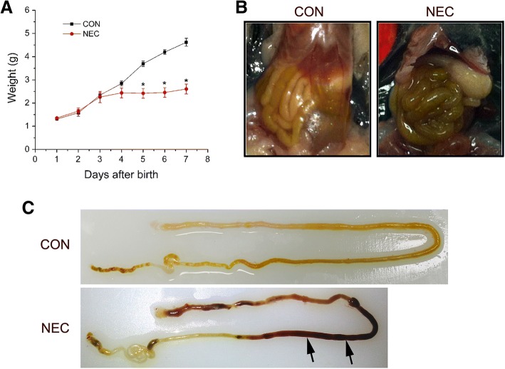
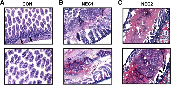
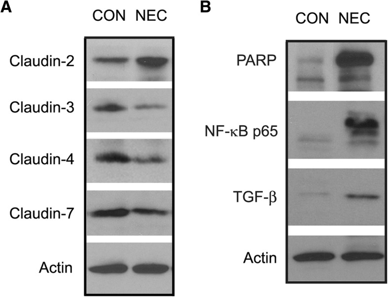
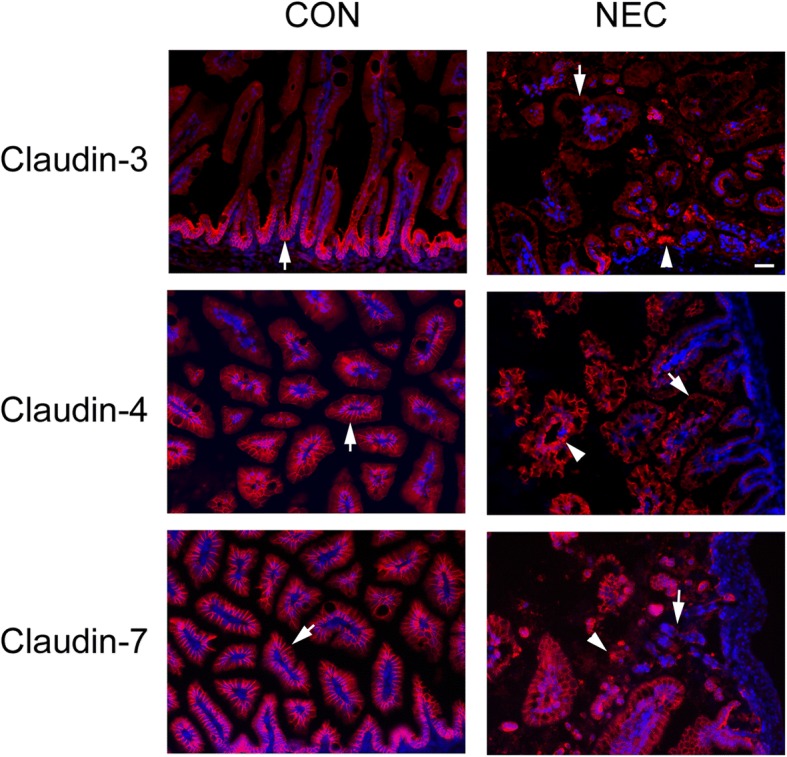
Methods: Three-day old C57BL/6 mouse pups were fed with Esbilac formula every 3 hours and then subjected to hypoxia twice daily followed by cold stress. Dam fed pups from the same litters served as controls. Pups were observed and sacrificed 96 hours after the treatments and intestines were removed for experiments. The successful induction of NEC was confirmed by histopathology. Changes in tight junction proteins in NEC intestines were studied by western blotting and immunofluorescent microscopy using specific protein markers. The gut leakage in NEC was visualized using biotin tracer molecules.

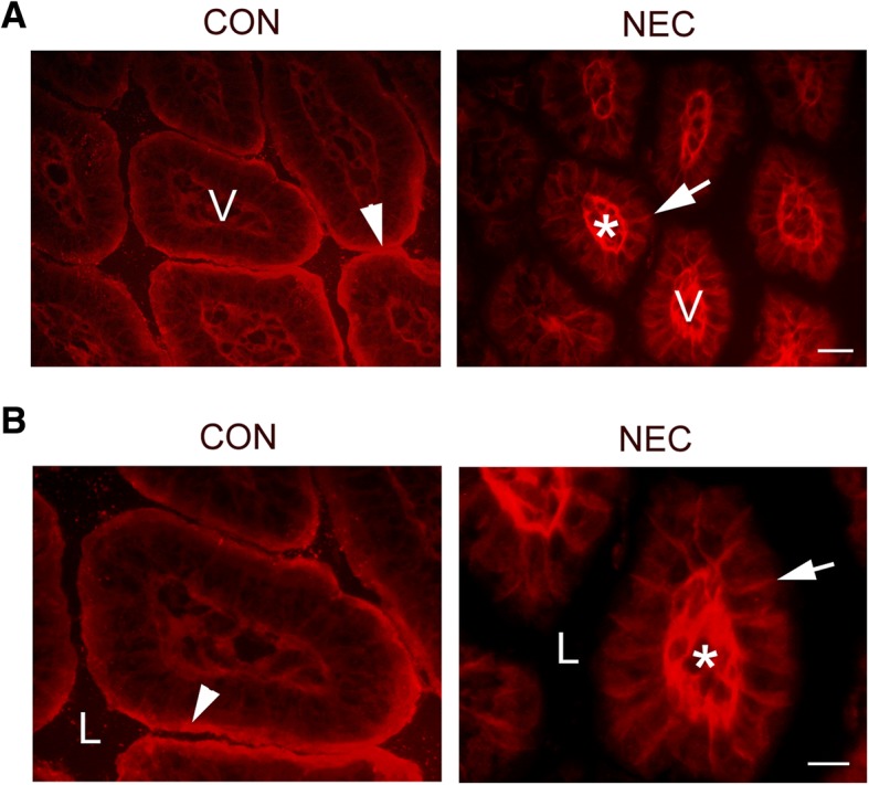
Results: Our study results demonstrate that we induced NEC in >50% of experimental pups, pups lost nearly 40% of weight and their intestines showed gross changes and microscopic changes associated with NEC. There were inflammatory changes with loss of tight junction barrier function and disruption of tight junction claudin proteins in the intestines of NEC mouse model. We have demonstrated for the first time that NEC intestines develop increased leakiness as visualized by biotin tracer leakage.

Conclusions: NEC leads to breakdown of epithelial barrier due to changes in tight junction proteins with increased leakiness which may explain the transmigration of microbes and microbial products from the gut lumen into the blood stream leading to sepsis like signs clinically witnessed.

Keywords: Biotin tracer molecules; Claudin proteins; Epithelial barrier function; Necrotizing enterocolitis; Tight junctions.


Conflict of interest statement

Ethics approval and consent to participate

Our animal study protocol was approved by the Animal Care and Use Committee of East Carolina University (AUP#A182). We performed the animal experiments according to the guidelines of East Carolina University and the National Institute of Health on animal care and use.

Consent for publication

Not applicable.

Competing interests

The authors declare that they have no competing interests.

Publisher’s Note

Springer Nature remains neutral with regard to jurisdictional claims in published maps and institutional affiliations.

Figures

Fig. 1
Fig. 1
Establishment of necrotizing enterocolitis (NEC) mouse model. a: Weight measurement of dam-fed (CON) or NEC-treated (NEC) pups. The treatment started at day 3 and was continued for 4 days. The data represent means ± s.e.m. from three independent experiments. Student’s t-test was used for statistical analysis. A P value of < 0.05 was considered significant (*). N=10 pups for each group. b: Comparison of control and NEC intestines in situ at the end of the experiments. The NEC intestine showed the swollen and discolored appearance. c: The control and NEC intestines after removal from the body. The NEC intestine displayed the severe hemorrhage as indicated by arrows
Fig. 2
Fig. 2
H& E staining of control and NEC intestines. a: Tissue sections from representative control intestines shows the normal villous structure with intact crypt region (arrows). b and c: Tissue sections from representative NEC intestinal samples. The induction of NEC led to stunting of villi and disrupted villous structure with sloughing of epithelial cells into the lumen (L). The hemorrhage is evident in both B and C NEC intestinal samples. Original magnification: 200×
Fig. 3
Fig. 3
Protein expression levels of control and NEC intestines. a: Representative western blot membrane showing claudin-2, -3, -4 and -7 expression levels in control (CON) and NEC intestines. Intestinal tissues from 7-day control and NEC pups were collected at the end of experiments. Tissue lysates were solubilized in RIPA buffer and subjected to western blotting. b: PARP, NF-κB and TGF-β expression levels in control (CON) and NEC intestines. A total of 30 μg protein for each sample were loaded on the SDS-polyacrylamide gel. Membranes were blotted against specific antibodies. Actin served as a loading control. Three independent experiments were performed
Fig. 4
Fig. 4
Immuno-localization of claudin proteins in control and NEC intestines. Representative images of intestines from 7-day control and NEC pups were dissected from the body and frozen in liquid nitrogen. Frozen sections were immunostained with anti-claudin-3, or -4, or -7 antibodies and detected by Cy3-conjugated secondary antibody. Nuclei were stained with DAPI (blue). The claudin signals were localized at cell-cell contact area as indicated by arrows in controls (CON). Arrows were pointed to the reduced signals in NEC samples and arrowheads in NEC showed the aggregated staining pattern. Scale bar: 50 μm
Fig. 5
Fig. 5
Biotin permeability assays in control and NEC intestines. a: Sulfo-NHS-LC-Biotin was injected into the intestinal lumen of 7-day old control (CON) or NEC pups. The tissue sections were stained with Texas red-conjugated streptavidin. Biotin was mainly kept at the epithelial surface of control intestines (arrowhead) due to the intact epithelial barriers. No barrier leakage was observed in control intestines. In contrast, the leakage was clearly detected in NEC intestines. Biotin stained the intercellular space (arrow) and connective tissue (Asterisk). V: Villus. b: The higher magnification of the images. L: Lumen. Scale bar: 50 μm in A and 20 μm in B

References

    1. Neu J, Walker WA. Necrotizing enterocolitis. N Engl J Med. 2011;364:255–264. doi: 10.1056/NEJMra1005408. - DOI - PMC - PubMed
    1. Pike K, Brocklehurst P, Jones D, Kenyon S, Salt A, Taylor D, et al. Outcomes at 7 years for babies who developed neonatal necrotising enterocolitis: the ORACLE Children Study. Arch Dis Child Fetal Neonatal Ed. 2012;97:F318–F322. doi: 10.1136/fetalneonatal-2011-300244. - DOI - PubMed
    1. Lin PW, Nasr TR, Stoll BJ. Necrotizing enterocolitis: recent scientific advances in pathophysiology and prevention. Semin Perinatol. 2008;32:70–82. doi: 10.1053/j.semperi.2008.01.004. - DOI - PubMed
    1. Gordon P, Christensen R, Weitkamp JH, Maheshwari A. Mapping the New World of Necrotizing Enterocolitis (NEC): Review and Opinion. EJ Neonatol Res. 2012;2:145–172. - PMC - PubMed
    1. Berseth CL. Gestational evolution of small intestine motility in preterm and term infants. J Pediatr. 1989;115:646–651. doi: 10.1016/S0022-3476(89)80302-6. - DOI - PubMed

Publication types

LinkOut - more resources