

Enhanced tendon-bone healing with acidic fibroblast growth factor delivered in collagen in a rabbit anterior cruciate ligament reconstruction model
- PMID: 30482233
- PMCID: PMC6260728
- DOI: 10.1186/s13018-018-0984-x
Enhanced tendon-bone healing with acidic fibroblast growth factor delivered in collagen in a rabbit anterior cruciate ligament reconstruction model
Abstract
Background: The objective of the present study was to investigate the effectiveness of acidic fibroblast growth factor delivered in collagen (aFGF/collagen) for promoting tendon-bone interface healing after anterior cruciate ligament (ACL) reconstruction in rabbits.
Methods: ACL reconstructions were performed in the right hind limbs of New Zealand rabbits. Each left long digital extensor tendon was harvested as an autograft, and collagen incorporating different concentrations of aFGF or same amount of collagen alone was applied at the tendon-bone interface after ACL reconstruction. The control group underwent ACL reconstruction only. There were high and low aFGF/collagen groups, collagen alone group, and control group (n = 21 rabbits per group). Histological and biomechanical analyses were performed at 4, 8, and 12 weeks postoperatively to evaluate the effect of aFGF/collagen on tendon-bone interface healing.
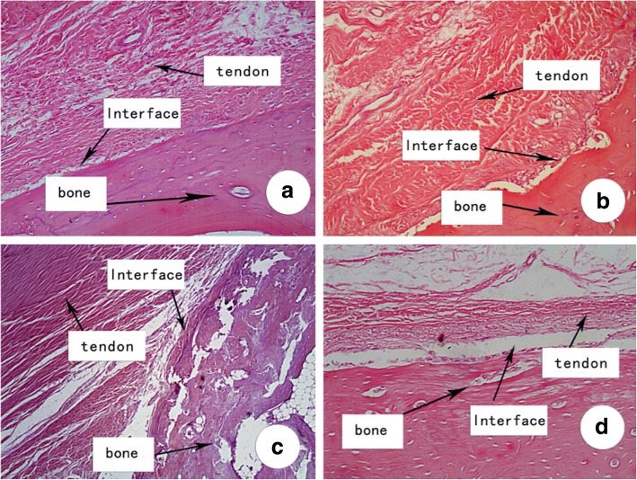
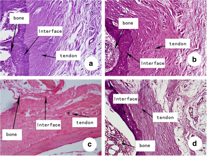
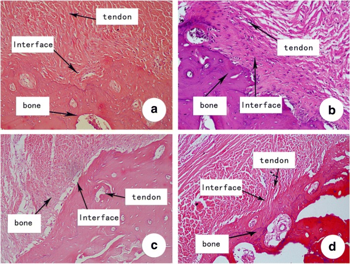
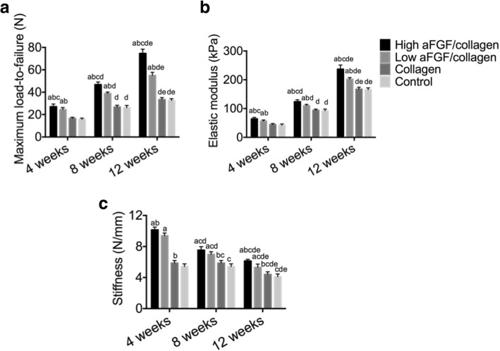
Results: Results of biomechanical tests showed that at both 8 and 12 weeks postoperatively, the elastic modulus and stiffness in both the high and low aFGF/collagen treatment groups were significantly higher than those in the control group and collagen alone group, with that in the high aFGF/collagen concentration group being the highest. Histological analysis showed that at 8 weeks, tightly organized Sharpey-like fibers were observed in both aFGF/collagen groups with new bone growth into the tendon in the high concentration group. At 12 weeks postoperatively, a fibrocartilage transition zone was observed in the bone tunnels in both aFGF/collagen groups, especially in the high aFGF/collagen group.
Conclusion: Application of the aFGF/collagen composite could enhance early healing at the tendon-bone interface after ACL reconstruction, especially with the use of a high aFGF/collagen concentration.
Keywords: Anterior cruciate ligament; Reconstruction; Tendon–bone healing; aFGF.
Conflict of interest statement
Ethics approval and consent to participate
This double-blinded study was approved by the Institutional Animal Care and Use committee at The Fourth Affiliated Hospital of Harbin Medical University and performed under the guidelines for the care and use of animals in research.
Consent for publication
Not applicable
Competing interests
The authors declare that they have no competing interests.
Publisher’s Note
Springer Nature remains neutral with regard to jurisdictional claims in published maps and institutional affiliations.
Figures
References
-
- Bourque WT, Gross M, Hall BK. Expression of four growth factors during fracture repair. Int J Dev Biol. 1993;37:573–579. - PubMed
-
- de Girolamo L, Galliera E, Volpi P, Denti M, Dogliotti G, Quaglia A, Cabitza P, Corsi Romanelli MM, Randelli P. Why menisci show higher healing rate when repaired during ACL reconstruction? Growth factors release can be the explanation. Knee Surg Sports Traumatol Arthrosc. 2015;23:90–96. doi: 10.1007/s00167-013-2712-8. - DOI - PubMed
MeSH terms
Substances
Grants and funding
LinkOut - more resources
Full Text Sources
Medical
