Review
. 2018:159:3-26.
doi: 10.1016/B978-0-444-63916-5.00001-X.

Sensorimotor anatomy of gait, balance, and falls

Affiliations
Review

Sensorimotor anatomy of gait, balance, and falls

Colum D MacKinnon. Handb Clin Neurol. 2018.

Abstract

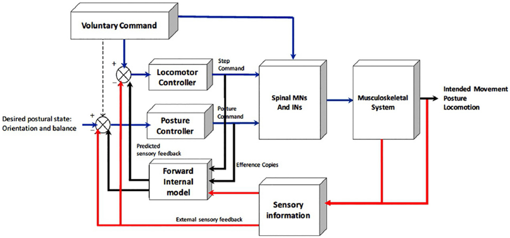
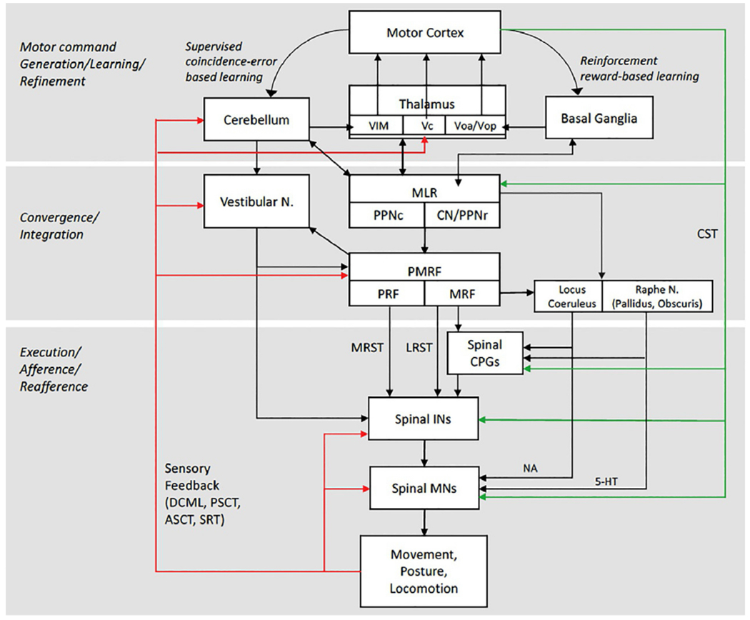
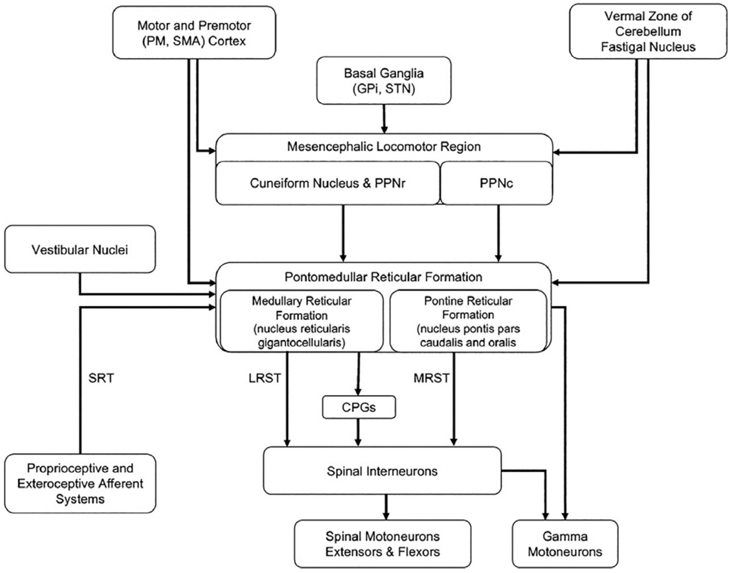
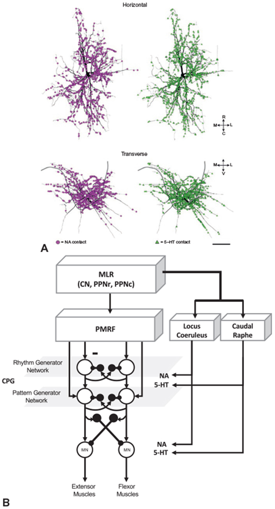
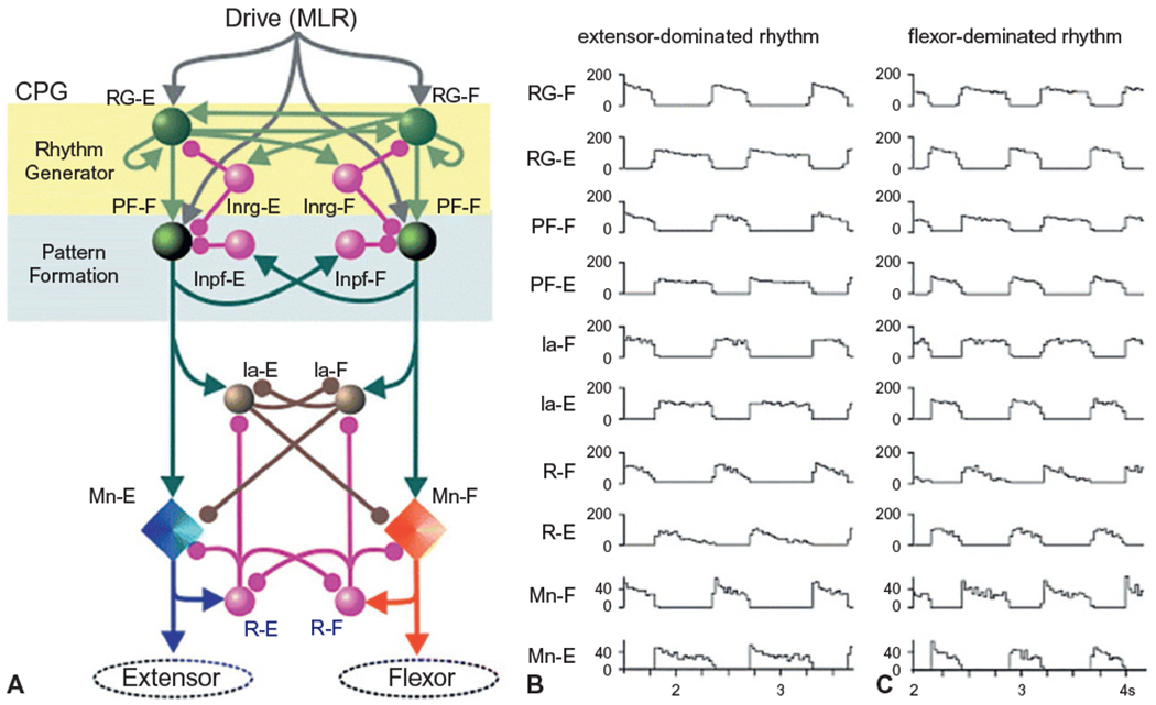
The review demonstrates that control of posture and locomotion is provided by systems across the caudal-to-rostral extent of the neuraxis. A common feature of the neuroanatomic organization of the postural and locomotor control systems is the presence of key nodes for convergent input of multisensory feedback in conjunction with efferent copies of the motor command. These nodes include the vestibular and reticular nuclei and interneurons in the intermediate zone of the spinal cord (Rexed's laminae VI-VIII). This organization provides both spatial and temporal coordination of the various goals of the system and ensures that the large repertoire of voluntary movements is appropriately coupled to either anticipatory or reactive postural adjustments that ensure stability and provide the framework to support the intended action. Redundancies in the system allow adaptation and compensation when sensory modalities are impaired. These alterations in behavior are learned through reward- and error-based learning processes implemented through basal ganglia and cerebellar pathways respectively. However, neurodegenerative processes or lesions of these systems can greatly compromise the capacity to sufficiently adapt and sometimes leads to maladaptive changes that impair movement control. When these impairments occur, the risk of falls can be significantly increased and interventions are required to reduce morbidity.

Keywords: balance; gait; locomotion; neuroanatomy; posture.


Figures

Fig. 1.1.
Fig. 1.1.
General model of the organization of the control of posture, locomotion, and voluntary movement. This model emphasizes that the voluntary command for movement (e.g., step initiation) must be coupled with parallel commands to a locomotor and postural control network. Efference copies of the locomotor and postural commands are relayed to a forward internal model (internal body schema), which, in conjunction with sensory information, is used to compare the desired postural/movement state with the perceived postural/movement state. Differences are used to update the locomotor and postural controller commands. INs, interneurons; MNs, motoneurons. (Adapted from Mille et al. (2012).)
Fig. 1.2.
Fig. 1.2.
Model of the core components of the nervous system, and their connectivity, that contribute to the control of balance, vertical support, posture, and locomotion and their integration with voluntary motor commands. 5-HT, serotonin; ASCT, anterior spinocerebellar tract; CPGs, central pattern generators; CST, corticospinal tract; DCML, dorsal column medial lemniscal tract; INs, interneurons; LRST, lateral (medullary) reticulospinal tract; MLR, mesencephalic locomotor region; MNs, motoneurons, MRF, medullary reticular formation; MRST, medial (pontine) reticulospinal tract; NA, noradrenaline (norepinephrine); PMRF, pontomedullary reticular formation; PPNc, caudal region of pedunculopontine nucleus; PPNr, rostral region of pedunculopontine nucleus; PRF, pontine reticular formation; PSCT, posterior spinocerebellar tract; SRT, spinoreticular tract; Vc, ventrocaudal nucleus of the thalamus; VIM, ventral intermediate nucleus of the thalamus; Voa/Vop, ventralis oralis anterior and ventralis oralis posterior of the thalamus.
Fig. 1.3.
Fig. 1.3.
(A) Diagram of a biologic muscle spindle and a model of spindle structure. A muscle spindle consists of three types of intrafusal fibers that receive fusimotor inputs (gamma static and dynamic) while giving rise to primary (Ia) and secondary (II) afferents. (B) Model of spindle output during 6-mm whole-muscle ramp stretches. Primary afferent responses at two different velocities (5 and 70 mm/s) are presented either in the absence of fusimotor stimulation (no gamma), with dynamic fusimotor stimulation at 70 pps, or during constant static fusimotor stimulation at 70 pps. Solid thin lines represent model output; experimental data are shown as dots. (C) Examples of the pattern of termination of single group Ia, group Ib, and group II muscle afferents from the triceps surae muscle in the cat. ((A) and (B) adapted from Mileusnic MP, Brown IE, Lan N, et al. (2006) Mathematical models of proprioceptors. I. Control and transduction in the muscle spindle. J Neurophysiol 96: 1772–1788, with permission from the American Physiological Society; (C) reproduced from Brown (1982) The dorsal horn of the spinal cord. Q J Exp Physiol 67: 193–212, with permission from John Wiley.)
Fig. 1.4.
Fig. 1.4.
Primary ascending sensory pathways that contribute to the control of posture, balance, and locomotion. (Reproduced from Parent A (1996) Carpenter’s human neuroanatomy, 9th edn. Baltimore, MD: Williams and Wilkins, with permission from Wolters Kluwer.)
Fig. 1.5.
Fig. 1.5.
(A) The vestibular apparatus and a simplified summary of its outputs and functions with respect to the control of posture and balance. (B) Summary of the structure, afferents, and efferents of the hair cells. (C) Model of the response of vestibular nucleus neurons to passive pure linear, pure rotational, and combined motion of the head. The bottom three rows show the changes in firing rates of a vestibular neuron that receives input from the semicircular canals, otolith, or combined input from both. RST, reticulospinal tract; VOR, vestibulo-ocular reflex; VST, vestibulospinal tract. ((A) Adapted with permission from Cullen KE (2012) The vestibular system: multimodal integration and encoding of self-motion for motor control. Trends Neurosci 35: 185–196. (B) Reproduced with permission from Parent A (1996) Carpenter’s human neuroanatomy, 9th edn. Baltimore, MD: Williams and Wilkins. (C) Reproduced from Carriot et al. (2015).)
Fig. 1.6.
Fig. 1.6.
Summary of the cortical (A) and subcortical (B) pathways linking the visual system input structures and connections of the visual system to structures that control movement and posture. RST, reticulospinal tract; VST, vestibulospinal tract.
Fig. 1.7.
Fig. 1.7.
Primary descending motor pathways that contribute to the control of posture, balance, and locomotion. (Reproduced from Parent A (1996) Carpenter’s human neuroanatomy, 9th edn. Baltimore, MD: Williams and Wilkins, with permission from Wolters Kluwer.)
Fig. 1.8.
Fig. 1.8.
Summary of the input and output pathways of the vestibular nucleus. VST, vestibulospinal tract. (Adapted from McCall AA, Miller DM, Yates BJ (2017) Descending influences on vestibulospinal and vestibulosympathetic reflexes. Front Neurol 8: 112.)
Fig. 1.9.
Fig. 1.9.
Summary of the input and output pathways to the mesencephalic locomotor region and pontomedullary reticular formation. CPGs, central pattern generators; GPi, globus pallidus internus; LRST, lateral (medullary) reticulospinal tract; MRST, medial (pontine) reticulospinal tract; PM, premotor cortex; PPNc, caudal region of pedunculopontine nucleus; PPNr, rostral region of pedunculopontine nucleus; SMA, supplementary motor area; SRT, spinoreticular tract; STN, subthalamic nucleus.
Fig. 1.10.
Fig. 1.10.
(A) Reconstruction of a spinal motoneuron showing the location and density of serotonergic (5-HT, green) and noradrenergic (NA, purple) inputs to the dendrites. (B) Summary of pathways mediating neuromodulatory (NA and 5-HT) inputs to the central pattern generator (CPG) network and spinal motoneurons from the brainstem. CN, cranial nerve; MLR, mesencephalic locomotor region; MN, motoneuron; PMRF, pontomedullary reticular formation; PPNc, caudal region of pedunculopontine nucleus; PPNr, rostral region of pedunculopontine nucleus. ((A) Reproduced from Montague SJ, Fenrich KK, Mayer-Macaulay C, et al. (2013) Nonuniform distribution of contacts from noradrenergic and serotonergic boutons on the dendrites of cat splenius motoneurons. J Comp Neurol 521: 638–656, with permission from John Wiley.)
Fig. 1.11.
Fig. 1.11.
(A) Model of a two-level central pattern generator (CPG) network. The CPG structure is organized into a layer of interneurons that control rhythm generation and a layer that controls pattern generation. Note the reciprocal connections between the flexors and extensors. Sensory feedback is transmitted to all layers of the network (not shown). (B and C) Locomotion patterns generated by the two-level model. Note the alternating activities of the flexor and extensor rhythm- (RG) and pattern- (PF) generating networks, flexor and extensor inhibitory interneurons (Ia) and Renshaw cells (R), and extensor spinal motoneuron activity (Mn). In panel B, the mesencephalic locomotor region drive to RG-E is larger than to RG-F and results in a rhythm with a longer duration extensor phase. Panel C demonstrates the rhythm generated when RG-F drive is larger than RG-E. (Reproduced from Rybak et al. (2006).)

Similar articles

Cited by

References

    1. Abzug C, Peterson BW (1973). Antidromic stimulation in the ponto-medullary reticular formation of local axon branches of contralateral vestibular neurons. Brain Res 64: 407–413. - PubMed
    1. Abzug C, Maeda M, Peterson BW et al. (1974). Cervical branching of lumbar vestibulospinal axons. J Physiol 243: 499–522. - PMC - PubMed
    1. Albin RL, Young AB, Penney JB (1989). The functional anatomy of basal ganglia disorders. Trends Neurosci 12: 366–375. - PubMed
    1. Baker SN (2011). The primate reticulospinal tract, hand function and functional recovery. J Physiol 589: 5603–5612. - PMC - PubMed
    1. Bannatyne BA, Edgley SA, Hammar I et al. (2006). Differential projections of excitatory and inhibitory dorsal horn interneurons relaying information from group II muscle afferents in the cat spinal cord. J Neurosci 26: 2871–2880. - PMC - PubMed