Review
. 2019 Feb;29(2):117-134.
doi: 10.1016/j.tcb.2018.09.007. Epub 2018 Oct 25.

Posing the APC/C E3 Ubiquitin Ligase to Orchestrate Cell Division

Affiliations
Review

Posing the APC/C E3 Ubiquitin Ligase to Orchestrate Cell Division

Edmond R Watson et al. Trends Cell Biol. 2019 Feb.

Abstract

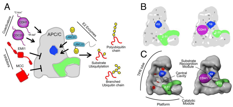
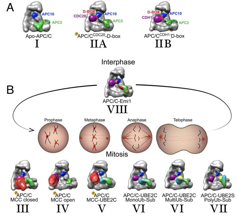
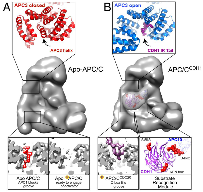
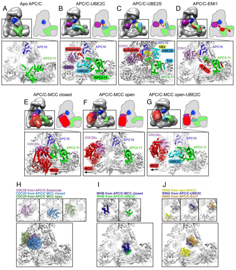
The anaphase promoting complex/cyclosome (APC/C) E3 ligase controls mitosis and nonmitotic pathways through interactions with proteins that coordinate ubiquitylation. Since the discovery that the catalytic subunits of APC/C are conformationally dynamic cullin and RING proteins, many unexpected and intricate regulatory mechanisms have emerged. Here, we review structural knowledge of this regulation, focusing on: (i) coactivators, E2 ubiquitin (Ub)-conjugating enzymes, and inhibitors engage or influence multiple sites on APC/C including the cullin-RING catalytic core; and (ii) the outcomes of these interactions rely on mobility of coactivators and cullin-RING domains, which permits distinct conformations specifying different functions. Thus, APC/C is not simply an interaction hub, but is instead a dynamic, multifunctional molecular machine whose structure is remodeled by binding partners to achieve temporal ubiquitylation regulating cell division.

Keywords: E3 ligase; anaphase promoting complex/cyclosome; cell division; cryo-electron microscopy; mitosis; ubiquitin.


Figures

Figure 1:
Figure 1:. Overall assembly of APC/C from cartoon-like views of APC/C from cryo EM maps low-pass filtered to 30 Å resolution to provide general topological insights.
A, Schematic of APC/C binding partners and regulators. Coactivators CDC20 and CDH1 (purple) bind substrates and conformationally activate the APC2-APC11 catalytic core; inhibitors EMI1 and MCC block activity in interphase and prior to anaphase, respectively; when liberated from inhibitors, the E2 enzymes UBE2C and UBE2S link Ub to substrates or substrate-linked Ubs, respectively, to generate products with various numbers and topologies of Ub modifications. B, 2D view of apo APC/C and APC/CCDH1 bound to a substrate peptide, showing positions of coactivator (CDH1, purple), a D-box motif sandwiched between the CDH1 β-propeller and APC10 (blue), and repositioning of APC2-APC11 (green) upon coactivator binding. Locations of other subunits are indicated by numbers, e.g. 1 refers to APC1. C, 3D views of structures in A, highlighting the TPR lobe, Platform, Catalytic module, central cavity, and four TPR grooves (red) that can bind coactivator, APC10, and MCC IR-tails and C-boxes.
Figure 2:
Figure 2:. Coactivator and catalytic core conformations in APC/C assemblies across the cell cycle from cartoon-like views cryo EM maps low-pass filtered to 30 Å resolution to provide general topological insights.
A, Apo and coactivator bound APC/C, which are the platforms for complexes formed throughout the cell cycle. From left to right, Conformation I observed in apo APC/C, with APC2-APC11 (green) catalytic core “down” and the C-terminal catalytic domain (C) autoinhibited through interactions with the platform. Conformation II, coactivator (purple) recruits a D-box substrate along with APC10 (navy), and results in shift of the catalytic core into an active, mobile “up” position. Presumably due to mobility, the catalytic core is lower resolution in EM maps of coactivator-bound APC/C, but is relatively higher occupancy for the complex of phosphorylated APC/CCDC20 bound to a D-box peptide (IIA) than for the corresponding complex with APC/CCDH1 (IIB). B, APC/C complexes with coactivators, inhibitors and E2s across the cell cycle. Prior to correct chromosome alignment on the mitotic spindle, MCC inhibits APC/CCDC20 in a closed configuration (Conformation III), where MCC (red) captures both CDC20A (purple) and the catalytic core (green) and fills the central cavity, and in an open conformation with MCC swung out of the central cavity and the catalytic core free to bind UBE2C (Conformation IV). When chromosomes are properly aligned on the spindle and cells are prepared for anaphase, MCC is ubiquitylated as a prelude to its dissociation from APC/CCDC20, by APC2-APC11 recruiting, activating, and positioning the E2 enzyme UBE2C (cyan) adjacent to MCC (Conformation V). Subsequently, during mitosis, coactivator bound APC/C recruits a variety of substrates -including a ubiquitylated (yellow) substrate - for UBE2C to place additional Ubs to be placed onto the substrate (Conformation VI). In Conformation VII, APC2 coordinates with UBE2S (cyan) at a site distal from the catalytic core, the RING domain of APC11 harbors an acceptor Ub for Lys-11 polyubiquitination (yellow). Conformation VIII, during interphase substrate recognition module and the catalytic core are blocked by multiple domains of EMI1 (red), which allows accumulation of cyclins to ultimately result in CDH1 phosphorylation, and resetting APC/C for interphase and another cell cycle.
Figure 3:
Figure 3:. Conformational activation of APC/C for coactivator and associated substrate binding.
A, Conformations of TPR subunits restricting coactivator binding. Middle, high resolution EM map of apo-APC/C, low-pass filtered to 30 Å to provide a global view of the IR-tail and C-box binding sites within APC/C. Top, crystal structure showing that in the absence of a coactivator’s IR tail peptide, APC3’s C-terminal domain (red) adopts a “closed” conformation where C-terminal helices of APC3 itself (indicated with arrow) are rearranged to occupy the IR tail-binding groove. Bottom, high resolution EM density of apo-APC/C. Left, the C-terminal TPR groove of one APC8 protomer (grey EM map) houses an autoinhibitory element from APC1 (red) that blocks CDC20 recruitment to unphosphorylated APC/C. Upon phosphorylation of this APC1 loop prevents its binding to APC8, freeing the APC8 groove (right), thereby allowing binding of CDC20 and cell cycle progression. B, Coactivator-bound conformations of TPR subunits and substrate-binding to coactivator. Middle, EM map of APC/CCDH1-substrate peptide complex, low-pass filtered to 30 Å to provide a global view of coactivator interactions, highlighting the location of the substrate-binding CDH1 α-propeller, and the IR-tail and C-box binding sites within APC/C. Top, “open”, active conformation of APC3’s C-terminal domain (blue) bound to the IR-tail from the C-terminus of CDH1 (magenta, indicated with arrow in same relative location as in panel A), visualized within high resolution EM density of an APC/CCDH1-EMI1 complex. Bottom, left, C-box binding as visualized in high-resolution EM map. When APC/C is phosphorylated, the APC8 pocket (grey EM density) can bind an N-terminal C-box, as shown from CDC20 (purple EM density). Right, model of substrate recognition module (APC10, navy and CDH1 β-propeller domain, purple) bound to peptides with ABBA, D-box, and KEN-box motifs (red) based on superimposing structures harboring these motifs on EM data showing the relative positions of substrate-bound CDH1 and APC10.
Figure 4:
Figure 4:. Molecular insights into conformational changes in the APC/C substrate recognition module and catalytic core acros the cell cycle.
Cartoon-like representations of APC/C are complemented with similarly colored high-resolution models. A, Without a coactivator bound in apo-APC/C, the catalytic core (green) is in an inactive “down” conformation and APC10 (navy) does not recruit substrates. B, In structures representing substrate ubiquitylation, with APC/C-coactivator (here CDH1, purple) complexes bound to substrate (red) and UBE2C (cyan), the catalytic conformation is established by cullin-RING domains (green, specifically the RING domain of APC11 and the extreme C-terminal WHB domain of APC2) grasping discrete surfaces from the UBE2C~Ub (yellow in cartoon) intermediate. This places the active site proximal to substrate lysines. C, In a model for polyubiquitylation, from APC/C-coactivator (here CDH1, purple) complexes with a substrate model (substrate peptide in red, a Ub variant mimicking its linked Ub in yellow) and UBE2S (cyan), the acceptor Ub (yellow) whose Lys11 will become linked to the C-terminus of another Ub is recruited by an unprecedented surface from the APC11 RING domain in the catalytic core (green) and positioned adjacent to the active site in UBE2S. In a unique E2–E3 interaction, the C-terminal helices of UBE2S’s catalytic domain are recruited via helices in APC2’s helical bundle and α/β-domain, and a C-terminal tail unique to UBE2S among the E2s is recruited to a pocket between APC2 and APC4. The location of UBE2S at the periphery of the APC/C central cavity may accommodate extension of long polyUb chains. D, High resolution cryo EM data showed multiple elements from EMI1 (red) block substrate binding and ubiquitylation by APC/CCDH1 during interphase. EMI1 occupies the substrate recognition module, and the catalytic module by wrapping around the APC11 RING domain, and inserting its own tail sequence in the same groove that would otherwise bind the tail from UBE2S. E, In near atomic resolution cryo EM data for a “closed” configuration, association of the MCC (red) is seen inhibiting substrate binding and ubiquitylation by UBE2C by reorienting and enwrapping the APC/C-bound CDC20 molecule (CDC20A, purple), capturing the UBE2C-binding APC2 WHB domain (green), and filling the central cavity. F, In “open” configurations represented by EM data that were lower resolution presumably due to mobility, the CDC20A (purple)-MCC (red) subcomplex is rotated out of the APC/C central cavity, and the catalytic module is liberated and available to bind E2s. G, In a structural model of MCC ubiquitylation based on low resolution EM data and high resolution structures of the individual components, the CDC20A (purple)-MCC (red) subcomplex is in an “open” configuration, while the APC11 RING domain and APC2 WHB domain grasp UBE2C as in panel B, except to place the active site adjacent to CDC20-bound MCC rather than to a coactivator-bound substrate. H, Close-up views of different functional positions of the CDC20 substrate receptor, from complex with substrate as represented by a D-box peptide (purple), with MCC in the closed configuration (slate), and with MCC in the open configuration (olive), individually on top and superimposed below. I, Close-ups of different functional positions of APC2 cullin WHB domain, as bound to MCC in the closed configuration (navy) and to UBE2C (green), individually on top and superimposed below. J, Close-ups of different functional positions of APC11 RING domain, as autoinhibited in apo APC/C (yellow), when poised to ubiquitylate substrates by activating the UBE2C~Ub intermediate (blue), and inhibited by EMI1 (orange) individually on top and superimposed below.

Similar articles

Cited by

References

    1. Yamano H et al. (1996) The role of proteolysis in cell cycle progression in Schizosaccharomyces pombe. EMBO J 15 (19), 5268–79. - PMC - PubMed
    1. King RW et al. (1996) How proteolysis drives the cell cycle. Science 274 (5293), 1652–9. - PubMed
    1. King RW et al. (1995) A 20S complex containing CDC27 and CDC16 catalyzes the mitosis-specific conjugation of ubiquitin to cyclin B. Cell 81 (2), 279–88. - PubMed
    1. Sudakin V et al. (1995) The cyclosome, a large complex containing cyclin-selective ubiquitin ligase activity, targets cyclins for destruction at the end of mitosis. Mol Biol Cell 6 (2), 185–97. - PMC - PubMed
    1. Irniger S et al. (1995) Genes involved in sister chromatid separation are needed for B-type cyclin proteolysis in budding yeast. Cell 81 (2), 269–78. - PubMed

Publication types

MeSH terms

Substances