

Bile Acid 7α-Dehydroxylating Gut Bacteria Secrete Antibiotics that Inhibit Clostridium difficile: Role of Secondary Bile Acids
- PMID: 30482679
- PMCID: PMC6338514
- DOI: 10.1016/j.chembiol.2018.10.003
Bile Acid 7α-Dehydroxylating Gut Bacteria Secrete Antibiotics that Inhibit Clostridium difficile: Role of Secondary Bile Acids
Abstract
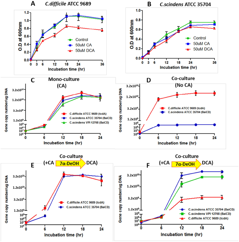
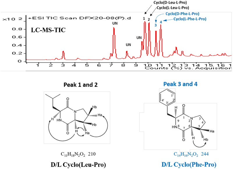
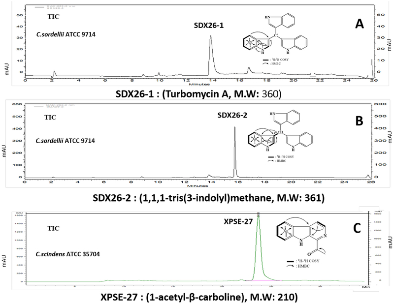
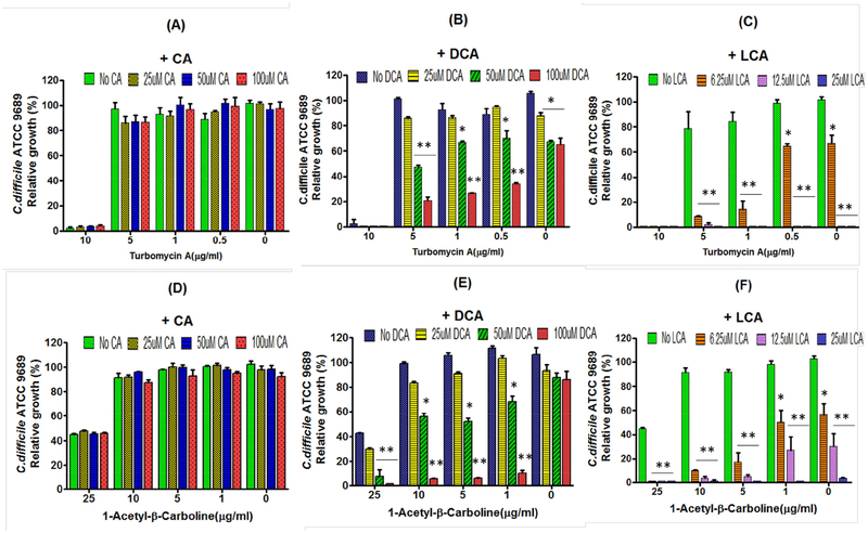
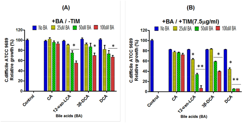
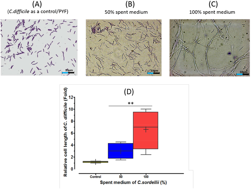
Clostridium scindens biotransforms primary bile acids into secondary bile acids, and is correlated with inhibition of Clostridium difficile growth in vivo. The aim of the current study was to determine how C. scindens regulates C. difficile growth in vitro and if these interactions might relate to the regulation of gut microbiome structure in vivo. The bile acid 7α-dehydroxylating gut bacteria, C. scindens and C. sordellii, were found to secrete the tryptophan-derived antibiotics, 1-acetyl-β-carboline and turbomycin A, respectively. Both antibiotics inhibited growth of C. difficile and other gut bacteria. The secondary bile acids, deoxycholic acid and lithocholic acid, but not cholic acid, enhanced the inhibitory activity of these antibiotics. These antibiotics appear to inhibit cell division of C. difficile. The results help explain how endogenously synthesized antibiotics and secondary bile acids may regulate C. difficile growth and the structure of the gut microbiome in health and disease.
Keywords: 1-acetyl-β-carboline; Clostridium difficile; Clostridium scindens; Clostridium sordellii; cyclic dipeptides; dysbiosis; gut microbiome; turbomycin A.
Published by Elsevier Ltd.
Conflict of interest statement
DECLARATION OF INTEREST
A patent application has been filed based on this study.
Figures
Comment in
-
Role of Bile in Infectious Disease: the Gall of 7α-Dehydroxylating Gut Bacteria.Cell Chem Biol. 2019 Jan 17;26(1):1-3. doi: 10.1016/j.chembiol.2018.12.010. Cell Chem Biol. 2019. PMID: 30658109 Free PMC article.
Similar articles
-
Metabolism of Oxo-Bile Acids and Characterization of Recombinant 12α-Hydroxysteroid Dehydrogenases from Bile Acid 7α-Dehydroxylating Human Gut Bacteria.Appl Environ Microbiol. 2018 May 1;84(10):e00235-18. doi: 10.1128/AEM.00235-18. Print 2018 May 15. Appl Environ Microbiol. 2018. PMID: 29549099 Free PMC article.
-
Functional Intestinal Bile Acid 7α-Dehydroxylation by Clostridium scindens Associated with Protection from Clostridium difficile Infection in a Gnotobiotic Mouse Model.Front Cell Infect Microbiol. 2016 Dec 20;6:191. doi: 10.3389/fcimb.2016.00191. eCollection 2016. Front Cell Infect Microbiol. 2016. PMID: 28066726 Free PMC article.
-
In vitro and in vivo characterization of Clostridium scindens bile acid transformations.Gut Microbes. 2019;10(4):481-503. doi: 10.1080/19490976.2018.1549420. Epub 2018 Dec 27. Gut Microbes. 2019. PMID: 30589376 Free PMC article.
-
Impact of microbial derived secondary bile acids on colonization resistance against Clostridium difficile in the gastrointestinal tract.Anaerobe. 2016 Oct;41:44-50. doi: 10.1016/j.anaerobe.2016.05.003. Epub 2016 May 7. Anaerobe. 2016. PMID: 27163871 Free PMC article. Review.
-
Interplay between Bile Acids and Intestinal Microbiota: Regulatory Mechanisms and Therapeutic Potential for Infections.Pathogens. 2024 Aug 20;13(8):702. doi: 10.3390/pathogens13080702. Pathogens. 2024. PMID: 39204302 Free PMC article. Review.
Cited by
-
Chemical Mechanisms of Colonization Resistance by the Gut Microbial Metabolome.ACS Chem Biol. 2020 May 15;15(5):1119-1126. doi: 10.1021/acschembio.9b00813. Epub 2020 Jan 14. ACS Chem Biol. 2020. PMID: 31895538 Free PMC article. Review.
-
The Mechanism of Important Components in Canine Fecal Microbiota Transplantation.Vet Sci. 2022 Dec 14;9(12):695. doi: 10.3390/vetsci9120695. Vet Sci. 2022. PMID: 36548856 Free PMC article. Review.
-
Role of Bile in Infectious Disease: the Gall of 7α-Dehydroxylating Gut Bacteria.Cell Chem Biol. 2019 Jan 17;26(1):1-3. doi: 10.1016/j.chembiol.2018.12.010. Cell Chem Biol. 2019. PMID: 30658109 Free PMC article.
-
Implications of Gut Microbiota in Complex Human Diseases.Int J Mol Sci. 2021 Nov 23;22(23):12661. doi: 10.3390/ijms222312661. Int J Mol Sci. 2021. PMID: 34884466 Free PMC article. Review.
-
Synthetic Microbiomes on the Rise-Application in Deciphering the Role of Microbes in Host Health and Disease.Nutrients. 2021 Nov 21;13(11):4173. doi: 10.3390/nu13114173. Nutrients. 2021. PMID: 34836426 Free PMC article. Review.
References
-
- Belin P, Moutiez M, Lautru S, Seguin J, Pernodet JL, Gondry M The nonribosomal synthesis of diketopiperazines in tRNA-dependent cyclodipeptide synthease pathways (2012). Nat Prod Rep. 29, 961–79. - PubMed
-
- Chen Y, Yang F, Lu H, Wang B, Chen Y Lei D, Wang Y, Zhu B, Li L (2011) Characterization of fecal microbial communities in patients with liver cirrhosis. Hepatology 54, 562–572. - PubMed
Publication types
MeSH terms
Substances
Grants and funding
LinkOut - more resources
Full Text Sources
Other Literature Sources
Medical
Molecular Biology Databases
