. 2019 Jan 15;79(2):320-327.
doi: 10.1158/0008-5472.CAN-18-1987. Epub 2018 Nov 27.

Ribosomal Lesions Promote Oncogenic Mutagenesis

Affiliations

Ribosomal Lesions Promote Oncogenic Mutagenesis

Sergey O Sulima et al. Cancer Res. .

Abstract

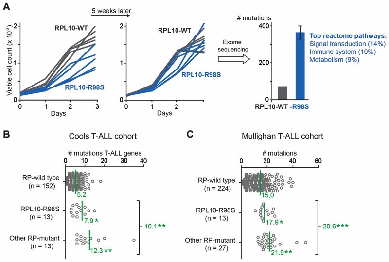
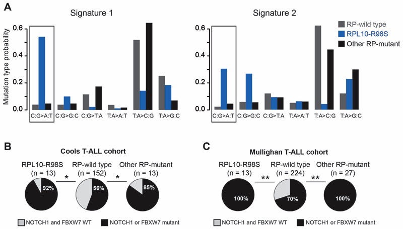
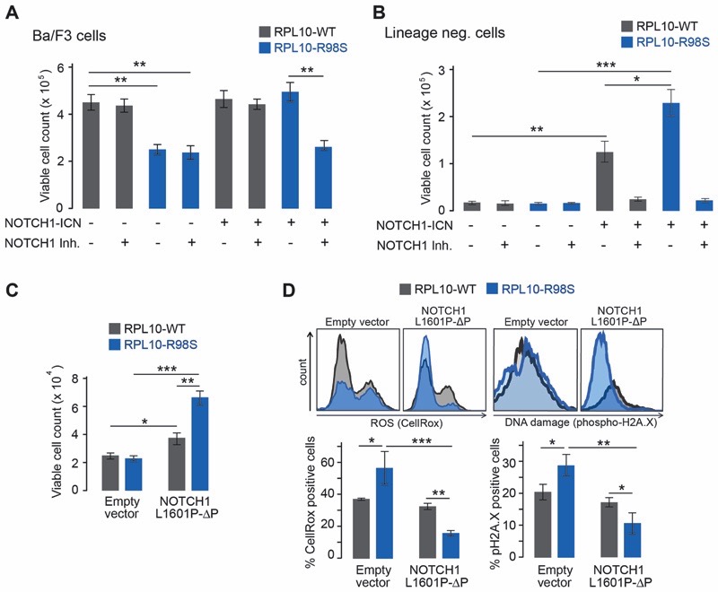
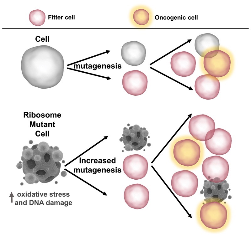
Ribosomopathies are congenital disorders caused by mutations in ribosomal proteins (RP) or assembly factors and are characterized by cellular hypoproliferation at an early stage. Paradoxically, many of these disorders have an elevated risk to progress to hyperproliferative cancer at a later stage. In addition, somatic RP mutations have recently been identified in various cancer types, for example, the recurrent RPL10-R98S mutation in T-cell acute lymphoblastic leukemia (T-ALL) and RPS15 mutations in chronic lymphocytic leukemia (CLL). We previously showed that RPL10-R98S promotes expression of oncogenes, but also induces a proliferative defect due to elevated oxidative stress. In this study, we demonstrate that this proliferation defect is eventually rescued by RPL10-R98S mouse lymphoid cells that acquire 5-fold more secondary mutations than RPL10-WT cells. The presence of RPL10-R98S and other RP mutations also correlated with a higher mutational load in patients with T-ALL, with an enrichment in NOTCH1-activating lesions. RPL10-R98S-associated cellular oxidative stress promoted DNA damage and impaired cell growth. Expression of NOTCH1 eliminated these phenotypes in RPL10-R98S cells, in part via downregulation of PKC-θ, with no effect on RPL10-WT cells. Patients with RP-mutant CLL also demonstrated a higher mutational burden, enriched for mutations that may diminish oxidative stress. We propose that oxidative stress due to ribosome dysfunction causes hypoproliferation and cellular insufficiency in ribosomopathies and RP-mutant cancer. This drives surviving cells, potentiated by genomic instability, to acquire rescuing mutations, which ultimately promote transition to hyperproliferation. SIGNIFICANCE: Ribosomal lesions cause oxidative stress and increase mutagenesis, promoting acquisition of rescuing mutations that stimulate proliferation.


Conflict of interest statement

The authors declare no competing financial interests

Figures

Figure 1
Figure 1. RP lesions increase the mutagenic burdenin T-ALL.
(A) Left and middle: growth curves of Ba/F3 cell clones expressing RPL10-R98S or RPL10-WT at different time points.Right: number of mutations as determined by exome sequencing on three cell clones (1 RPL10-WT and 2 RPL10-R98S) from the left panel of Figure 1A. The percentages in the reactome pathways correspond to the fraction of mutated genes in the RPL10-R98S clones belonging to the mentioned reactome pathway. (B)The number of mutations in genes known to be recurrently mutated in T-ALL (T-ALL genes) in RP-mutant vs. RP-WT T-ALL patient samples, mined from targeted resequencing data (11). The RPL10-R98S category includes patients with RPL10-R98S as the only RP mutation. The “other RP-mutant” category includespatients with mutations in RPL5, RPL22, or in multiple RPs including RPL10-R98S. Mean values are indicated by a green line and the corresponding green number. (C) The number of mutations in RP-mutant vs. RP-WT T-ALL, mined from patient whole exome data (12). The RPL10-R98S category includes patients with RPL10-R98S as the only RP mutation. The “other RP-mutant” category includes a patient with the RPL10-R98C mutation, a patient with the RPL10-Q123P mutation, and patients with mutations in RPL4, RPL5, RPL9, RPL11, RPL22, RPS3, RPS13, or in multiple RPs. Mean values are indicated by a green line. P-values were calculated using a T-test after accounting for equality of variances using an F-test (panels B and C). ***P ≤ 0.001, **P ≤ 0.01, *P ≤ 0.05.
Figure 2
Figure 2. RP-mutant T-ALL displays unique mutational signatures and enrichment in NOTCH1 pathway activating mutations
(A)Mutational signature analysis (13) performed on a T-ALL whole exome sequencing cohort (12). RPL10-R98S samples showed a significant differencefor signature 1 compared to RP-WT samples (P = 0.0044). The C:G>A:T transversion mutations, which mainly correspond to the type of mutations linked to oxidative DNA damage, are indicated by black rectangles. (B) Fraction of patients with mutations in NOTCH1 pathway genes in RP-mutant vs. RP-WT T-ALL, mined from targeted resequencing data (11).The RPL10-R98S category includes patients with RPL10-R98S as the only RP mutation. The “other RP-mutant” category includes patients with mutations in RPL5, RPL22, or in multiple RPs including RPL10-R98S. (C) Fraction of patients with mutations in NOTCH1 pathway genes in RP-mutant vs. RP-WT T-ALL, mined from whole exome data (12). The RPL10-R98S category includes patients with RPL10-R98S as the only RP mutation. The “other RP-mutant” category includes a patient with the RPL10-R98C mutation,a patient with the RPL10-Q123P mutation, and patients with mutations in RPL4, RPL5, RPL9, RPL11, RPL22, RPS3, RPS13, or in multiple RPs. A Wilcoxon rank sum test was performed in Panel A, and a two-tailed Fisher’s test in Panels B-C. **P ≤ 0.01, *P ≤ 0.05.
Figure 3
Figure 3. NOTCH1 rescues theproliferation and functional defectsof a RP-mutant T-ALL model
(A) Cell count of Ba/F3 clones expressing RPL10-R98S or RPL10-WT and NOTCH1-ICN or empty vector, with or without 10 µM of the NOTCH1 inhibitor (NOTCH1 Inh.) SAHM1. (B) Cell count of lin- cells in a serial replating assay expressing RPL10-R98S or RPL10-WT and NOTCH1-ICN or empty vector, with or without the NOTCH1 inhibitor (NOTCH1 Inh.) SAHM1. (C)Cell count of lin- cells cultured on cytokine-rich semi-solid media expressing RPL10-R98S or RPL10-WT, transduced with NOTCH1-L1601P-?PEST (NOTCH1-L1601P-ΔP) or empty vector. (D)Levels of ROS (CellRox) and DNA damage (phospho-H2A.X) in viable lin- cells expressing RPL10-R98S or RPL10-WT, transduced with NOTCH1-L1601P-ΔPEST (NOTCH1-L1601P-ΔP) or empty vector. A representative staining for each condition is depicted, along with bar graphs indicating the average values from at least 3 biological replicates. Error bars denote standard deviation. P-values were calculated using a T-test after accounting for equality of variances using an F-test. ***P ≤ 0.001, **P ≤ 0.01, *P ≤ 0.05. N = 3-6.
Figure 4
Figure 4. Model: the role of RP lesions in ribosome-defective disorders
By promoting oxidative stress and DNA damage, RP lesions provide a fertile ground for elevated mutagenesis. This increases the likelihood of acquiring disease-specific rescuing and/or cooperating lesions, such as mutations in the NOTCH1 and TP53 pathways in T-ALL and CLL, respectively.

Similar articles

Cited by

References

    1. Sulima SO, Hofman IJF, De Keersmaecker K, Dinman JD. How Ribosomes Translate Cancer. Cancer Discov. 2017;7:1069–1087. - PMC - PubMed
    1. Dameshek W. Riddle: what do aplastic anemia, paroxysmal nocturnal hemoglobinuria (PNH) and “hypoplastic” leukemia have in common? Blood. 1967;30:251–254. - PubMed
    1. De Keersmaecker K, Sulima SO, Dinman JD. Ribosomopathies and the paradox of cellular hypo- to hyperproliferation. Blood. 2015;125:1377–1382. - PMC - PubMed
    1. De Keersmaecker K, Atak ZK, Li N, Vicente C, Patchett S, Girardi T, et al. Exome sequencing identifies mutation in CNOT3 and ribosomal genes RPL5 and RPL10 in T-cell acute lymphoblastic leukemia. Nat Genet. 2012;45:186–190. - PMC - PubMed
    1. Sulima SO, Patchett S, Advani VM, De Keersmaecker K, Johnson AW, Dinman JD. Bypass of the pre-60S ribosomal quality control as a pathway to oncogenesis. Proc Natl Acad Sci USA. 2014;111:5640–5645. - PMC - PubMed

Publication types

MeSH terms