. 2019 Jan 18;294(3):816-826.
doi: 10.1074/jbc.RA117.000848. Epub 2018 Nov 27.

Sodium channel TRPM4 and sodium/calcium exchangers (NCX) cooperate in the control of Ca2+-induced mucin secretion from goblet cells

Affiliations

Sodium channel TRPM4 and sodium/calcium exchangers (NCX) cooperate in the control of Ca2+-induced mucin secretion from goblet cells

Gerard Cantero-Recasens et al. J Biol Chem. .

Abstract

Regulated mucin secretion is essential for the formation of the mucus layer that protects the underlying epithelial cells from foreign particles. Alterations in the quantity or quality of secreted mucins are therefore detrimental to airway and colon physiology. Based on various biochemical assays in several human cell lines, we report here that Na+/Ca2+ exchanger 2 (NCX2) works in conjunction with transient receptor potential cation channel subfamily M member 4 (TRPM4), and perhaps TRPM5, Na+ channels to control Ca2+-mediated secretion of both mucin 2 (MUC2) and MUC5AC from HT29-18N2 colonic cancer cells. Differentiated normal bronchial epithelial (NHBE) cells and tracheal cells from patients with cystic fibrosis (CFT1-LC3) expressed only TRPM4 and all three isoforms of NCXs. Blocking the activity of TRPM4 or NCX proteins abrogated MUC5AC secretion from NHBE and CFT1-LC3 cells. Altogether, our findings reveal that NCX and TRPM4/TRPM5 are both required for mucin secretion. We therefore propose that these two proteins could be potential pharmacological targets to control mucus-related pathologies such as cystic fibrosis.

Keywords: MUC2; MUC5AC; TRPM4; TRPM5; cystic fibrosis; goblet cell; mucin; secretion; sodium-calcium exchange; transient receptor potential channels (TRP channels).


Conflict of interest statement

The authors declare that they have no conflicts of interest with the contents of this article

Figures

Figure 1.
Figure 1.
TRPM4/TRPM5 and NCX2 are required for mucin secretion by HT29-18N2 colonic cancer cells. A, RNA levels of NCX1, NCX2, NCX3, TRPM4, TRPM5, MUC2, and MUC5AC in positive control cDNA (control cDNA for NCX1, NCX2, TRPM4; lungs cDNA for NCX3, TRPM5, and MUC5AC; and colon cDNA for MUC2; MTC cDNA Panels, Clontech Laboratories) and differentiated HT29-18N2 cDNA were analyzed by agarose gel. B, intracellular MUC5AC levels of control, TRPM4, TRPM5, TRPM4 + TRPM5, and NCX2 KD cells. Differentiated cells were lysed and analyzed by dot blot with anti-MUC5AC and anti-actin antibodies. Intensities of MUC5AC spots were normalized to the actin levels. Results are mean ± S.E. (n ≥ 3). C, intracellular MUC2 levels of differentiated control, TRPM4 KD, TRPM5 KD, TRPM4 + TRPM5 KD, and NCX2 KD cells. Cells were lysed and analyzed by dot blot with an anti-MUC2 and anti-actin antibody. Intensities of MUC2 spots were quantified using ImageJ and normalized to the actin levels. Results are mean ± S.E. (n = 3). D, control, TRPM4, TRPM5, TRPM4 + TRPM5, and NCX2 stable knockdown cells were starved and incubated for 30 min at 37 °C with 100 μm ATP or vehicle. Secreted MUC5AC was collected, processed for dot blot analysis with an anti-MUC5AC antibody, and quantified using ImageJ. The y axis represents relative values with respect to the values of untreated cells of each condition. Average values ± S.E. are plotted as bar graphs (n ≥ 3). Statistics are shown for 100 μm ATP condition. E, differentiated control, TRPM4 KD, TRPM5 KD, TRPM4 + TRPM5 KD, and NCX2 KD cells were incubated for 30 min at 37 °C with 100 μm ATP or vehicle. Secreted MUC2 was collected and analyzed by dot blot with an anti-MUC2 antibody and analyzed with ImageJ. The y axis of the plot represents relative values with respect to the values of untreated cells for each condition. Average values ± S.E. are plotted as bar graphs (n = 3). Statistical analyses are only shown for 100 μm ATP stimulation. F, control, TRPM4 KD, TRPM5 KD, TRPM4 + TRPM5 KD, and NCX2 KD cells were differentiated by starvation. After starvation, cells were processed for cytosolic washout, fixed, and permeabilized for analysis by immunofluorescence microscopy with anti-MUC5AC antibody (red), anti-MUC2 antibody (green), and DAPI (blue). Scale bar = 5 μm. Ctrl, control; Neg, reverse transcription without reverse transcriptase; Scr., scrambled shRNA; TRPM4/5 shRNA, TRPM4 + TRPM5 KD cells. *, p < 0.05; **, p < 0.01.
Figure 2.
Figure 2.
TRPM4/5 and NCX control ATP-mediated Ca2+ entry. A, HT29-18N2 cells were starved for 6 days and incubated for 30 min at 37 °C with 100 μm ATP or vehicle in the presence or absence of extracellular Ca2+. Secreted MUC5AC was collected and processed for dot blot analysis with an anti-MUC5AC antibody. The dot blots were quantified using ImageJ. The y axis represents relative values with respect to the values of vehicle-treated cells for each condition. Average values ± S.E. are plotted as bar graphs (n = 3). B, secreted medium from differentiated HT29-18N2 after treatment with 100 μm ATP or the vehicle in the presence or absence of extracellular Ca2+ was processed for dot blot analysis with an anti-MUC2 antibody. The y axis represents relative values with respect to the values of vehicle-treated cells for each condition. Average values ± S.E. are plotted as bar graphs (n = 3). C, time course of mean Ca2+ responses (normalized Fura-2/AM ratio) obtained in differentiated HT29 cells treated with 100 μm ATP (●) or 1 μm TG (○) (n > 3) in the absence (calcium release from internal stores) or presence (calcium entry) of 1.2 mm CaCl2. D, average AUC from the traces shown in C in the absence of Ca2+ (stores release, AUC for 8 min) or in the presence of extracellular Ca2+ (Ca2+ entry, AUC for 10 min). E, differentiated HT29-18N2 cells were incubated for 30 min at 37 °C with 100 μm ATP, 1 μm TG, or vehicle. Secreted MUC5AC was collected and processed for dot blot with an anti-MUC5AC antibody. The y axis represents relative values to vehicle condition. Results are average values ± S.E. (n = 3). F, time course of mean Ca2+ responses (normalized Fura-2/AM ratio) obtained in differentiated HT29-18N2 cells (Control, TRPM4 KD, TRPM5 KD, TRPM4 + TRPM5 KD, and NCX2 KD) treated with 100 μm ATP (n ≥ 3) in the absence (stores release) or presence (Ca2+ entry) of extracellular Ca2+. G, average AUC (5 min for stores release, 10 min for Ca2+ entry) obtained from traces shown in C. Abbreviations: Veh, vehicle; TRPM4/5 shRNA, TRPM4 + TRPM5 KD cells. **, p < 0.01.
Figure 3.
Figure 3.
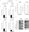
Differentiation of airway cells. A, schematic representation of airlift differentiation protocol. B, total RNA was extracted from undifferentiated (UND) and 21-day differentiated (DIF) NHBE and CFT1-LC3 cells. RNA levels of MUC5AC were monitored by qPCR (GAPDH was used as a reference gene). C, undifferentiated and 21-day differentiated NHBE and CFT1-LC3 cells were lysed and the lysates were analyzed by dot blot with an anti-MUC5AC antibody. The intensity of the spots was quantified using ImageJ and normalized to β-actin levels and to undifferentiated cells. Results are mean ± S.E. (n = 3). D, undifferentiated and 21-day differentiated NHBE and CFT1-LC3 cells were treated for 1 h at 37 °C with stimuli. Secreted MUC5AC was collected and analyzed by dot blot with an anti-MUC5AC antibody. The dot blots were quantified using ImageJ. Results are mean ± S.E. (n = 3). E, total RNA was extracted from 21-day differentiated NHBE and CFT1-LC3 cells. RNA levels of NCX1, NCX2, NCX3, TRPM4, TRPM5, and MUC5AC were analyzed by agarose gel electrophoresis. HPRT1 was used as a loading control. D21, day 21; Neg, negative; Ext., extraction. *, p < 0.05; **, p < 0.01.
Figure 4.
Figure 4.
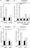
Involvement of NCXs in MUC5AC secretion by airway cells. A, differentiated NHBE cells were pretreated for 15 min at 37 °C with DMSO (Control) or 50 μm KB-R7943 (NCX inh.). After 15 min, a mixture containing 100 μm ATP + 10 ng/ml of IL-13 + 300 nm PMA, and the respective drug (DMSO or KB-R7943) was added to the cells and incubated for 1 h at 37 °C. Secreted MUC5AC was collected and processed for dot blot with an anti-MUC5AC antibody. The y axis represents relative values with respect to the values of the basal of each condition. Results are average values ± S.E. (n ≥ 3). B, differentiated CFT1-LC3 cells were pretreated for 15 min at 37 °C with DMSO (Control), 50 μm KB-R7943 (KB-R), 5 μm benzamil (Benz.), or 5 μm SN-6. After 15 min, a mixture containing 100 μm ATP + 10 ng/ml of IL-13 + 300 nm PMA, and the respective drug (DMSO, KB-R7943, benzamil, or SN-6) was added to the cells and incubated for 1 h at 37 °C. Secreted MUC5AC was collected and processed for dot blot with an anti-MUC5AC antibody. The y axis represents relative values with respect to the values of the basal of each condition. Results are average values ± S.E. (n ≥ 4). C, differentiated CFT1-LC3 cells were incubated for 1 h at 37 °C with DMSO (Control) or 10 μm 9-phenanthrol (TRPM4 inh.) in the presence or absence of stimuli (100 μm ATP + 10 ng/ml of IL-13 + 300 nm PMA). Secreted MUC5AC was collected and processed for dot blot with an anti-MUC5AC antibody. The y axis represents relative values with respect to the values of the basal for each condition. Results are average values ± S.E. (n ≥ 3). D, CFT1-LC3 differentiated cells were incubated for 1 h at 37 °C with a mixture containing 100 μm ATP + 10 ng/ml of IL-13 + 300 nm PMA or vehicle in the presence (w/Ca2+) or absence of Ca2+ (wo/Ca2+). Secreted MUC5AC was collected and processed for dot blot analysis with an anti-MUC5AC antibody. The dot blots were quantified using ImageJ. The y axis represents relative values with respect to the values of vehicle-treated cells for each condition. Average values ± S.E. are plotted as bar graphs (n = 3). NCX inh., NCX inhibitor KB-R7943; TRPM4 inh., TRPM4 inhibitor 9-phenanthrol. *, p < 0.05; **, p < 0.01.
Figure 5.
Figure 5.
A working model for MUC5AC secretion. A, baseline mucin secretion. In the absence of an external stimulus, TRPM4/TRPM5 are inactive. The secretion of mucin under these conditions is regulated by KChIP3 and intracellular calcium oscillations (7). B, stimulated mucin secretion. After stimulation with an exogenous stimulus, TRPM4/TRPM5 are active and permit sodium into the cell. This pool of Na+ triggers NCX to operate in the reverse mode whereby Na+ is pumped out and Ca2+ is imported into the cytoplasm. The local increase in Ca2+ by this procedure triggers mucin secretion. Rec, receptor; IP3R, inositol 1,4,5-trisphosphate receptor; Ext, extracellular; Cyt, cytosol.

Similar articles

Cited by

References

    1. Linden S. K., Sutton P., Karlsson N. G., Korolik V., and McGuckin M. A. (2008) Mucins in the mucosal barrier to infection. Mucosal Immunol. 1, 183–197 10.1074/jbc.RA117.001068 - DOI - PMC - PubMed
    1. Thornton D. J., Rousseau K., and McGuckin M. A. (2008) Structure and function of the polymeric mucins in airways mucus. Annu. Rev. Physiol. 70, 459–486 10.1074/jbc.RA117.001068 - DOI - PubMed
    1. Johansson M. E., Larsson J. M., and Hansson G. C. (2011) The two mucus layers of colon are organized by the MUC2 mucin, whereas the outer layer is a legislator of host-microbial interactions. Proc. Natl. Acad. Sci. U.S.A. 108, 4659–4665 10.1074/jbc.RA117.001068 - DOI - PMC - PubMed
    1. Rose M. C., and Voynow J. A. (2006) Respiratory tract mucin genes and mucin glycoproteins in health and disease. Physiol. Rev. 86, 245–278 10.1074/jbc.RA117.001068 - DOI - PubMed
    1. Adler K. B., Tuvim M. J., and Dickey B. F. (2013) Regulated mucin secretion from airway epithelial cells. Front. Endocrinol. 4, 129 10.3389/fendo.2013.00129 10.3389/fendo.2013.00129 - DOI - DOI - PMC - PubMed

Publication types

LinkOut - more resources