. 2018 Dec 11;115(50):E11857-E11863.
doi: 10.1073/pnas.1815410115. Epub 2018 Nov 27.

PYL8 mediates ABA perception in the root through non-cell-autonomous and ligand-stabilization-based mechanisms

Affiliations

PYL8 mediates ABA perception in the root through non-cell-autonomous and ligand-stabilization-based mechanisms

Borja Belda-Palazon et al. Proc Natl Acad Sci U S A. .

Abstract

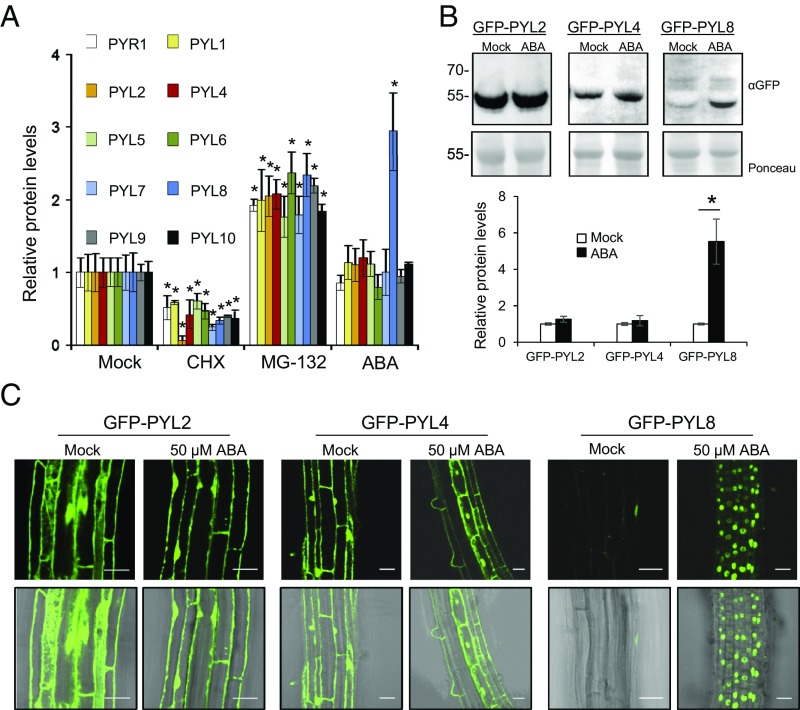
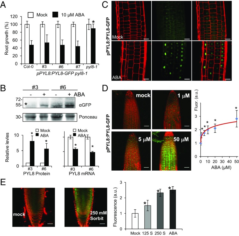
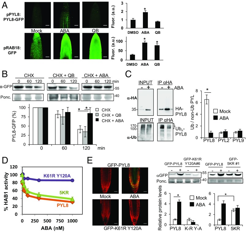
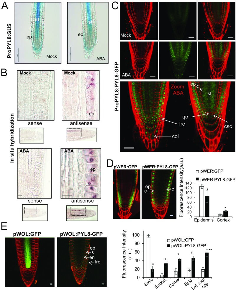
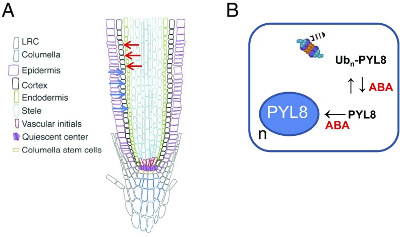
The phytohormone abscisic acid (ABA) plays a key role regulating root growth, root system architecture, and root adaptive responses, such as hydrotropism. The molecular and cellular mechanisms that regulate the action of core ABA signaling components in roots are not fully understood. ABA is perceived through receptors from the PYR/PYL/RCAR family and PP2C coreceptors. PYL8/RCAR3 plays a nonredundant role in regulating primary and lateral root growth. Here we demonstrate that ABA specifically stabilizes PYL8 compared with other ABA receptors and induces accumulation of PYL8 in root nuclei. This requires ABA perception by PYL8 and leads to diminished ubiquitination of PYL8 in roots. The ABA agonist quinabactin, which promotes root ABA signaling through dimeric receptors, fails to stabilize the monomeric receptor PYL8. Moreover, a PYL8 mutant unable to bind ABA and inhibit PP2C is not stabilized by the ligand, whereas a PYL85KR mutant is more stable than PYL8 at endogenous ABA concentrations. The PYL8 transcript was detected in the epidermis and stele of the root meristem; however, the PYL8 protein was also detected in adjacent tissues. Expression of PYL8 driven by tissue-specific promoters revealed movement to adjacent tissues. Hence both inter- and intracellular trafficking of PYL8 appears to occur in the root apical meristem. Our findings reveal a non-cell-autonomous mechanism for hormone receptors and help explain the nonredundant role of PYL8-mediated root ABA signaling.

Keywords: ABA; ABA biosensor; PYL8; non-cell-autonomous; root.


Conflict of interest statement

The authors declare no conflict of interest.

Figures

Fig. 1.
Fig. 1.
ABA treatment specifically increases PYL8 protein levels in seedlings. (A) Effect of CHX, MG132, or ABA treatment on protein levels of HA-tagged receptors. The 10-d-old seedlings expressing HA-tagged receptors were either mock or chemically treated with 50 μM CHX, MG132, or ABA for 6 h. Immunoblot analysis using anti-HA was performed to quantify protein levels. A single asterisk (*) indicates P < 0.05 (Student’s t test) compared with the corresponding mock-treated sample. (B) Effect of ABA treatment on GFP-PYL2, GFP-PYL4, and GFP-PYL8 protein levels. Seedlings expressing GFP-tagged PYL proteins were either mock- or 50 μM ABA-treated for 6 h. Immunoblot analysis using anti-GFP was performed to quantify protein levels. (C) ABA treatment leads to selective accumulation of GFP-PYL8 in the nucleus. CLSM analysis of the Arabidopsis root differentiation zone in lines expressing GFP-tagged PYL proteins that were either mock- or ABA-treated for 1 h. (Scale bars, 30 μm.)
Fig. 2.
Fig. 2.
ABA increases PYL8 protein levels in roots through a posttranscriptional mechanism. (A) PYL8-GFP complements the ABA-insensitive pyl8-1 phenotype. The 5-d-old seedlings germinated on Murashige and Skoog plates were transferred to new plates lacking or supplemented with 10 μM ABA, and quantification of root growth was performed after 10 d. Data are averages ±SD from three independent experiments (n = 20). A single asterisk (*) indicates P < 0.05 (Student’s t test) compared with Col-0 in the same assay conditions. (B) ABA treatment leads to accumulation of PYL8-GFP protein and down-regulation of PYL8-GFP mRNA in roots. The 10-d-old seedlings expressing PYL8-GFP were either mock- or 50 μM ABA-treated for 3 h and protein or RNA extracted from root tissue. Immunoblot analysis using anti-GFP was performed to quantify protein levels of PYL8-GFP (asterisk) in roots. A major 30-kDa root protein was used to normalize protein loading. qRT-PCR analyses were performed to quantify mRNA expression of PYL8-GFP. A single asterisk (*) indicates P < 0.05 (Student’s t test) compared with mock-treated samples. (C) ABA treatment leads to accumulation of PYL8-GFP in the nucleus. CLSM analysis of Arabidopsis root expressing ProPYL8:PYL8-GFP in the pyl8-1 background after mock or ABA treatment for 6 h. (Scale bars, 25 μm.) (D) Dose–response analysis of PYL8-GFP accumulation in response to treatment with the indicated ABA concentrations for 6 h. Fluorescence was quantified in arbitrary units (a.u.) using images acquired by CLSM. (E) Accumulation of PYL8-GFP after 250 mM sorbitol treatment. Fluorescence was measured after treatment with 125 or 250 mM sorbitol (S) or 50 μM ABA for 3 h. (Scale bars, 30 μm.) A single asterisk indicates P < 0.05 (Student’s t test) compared with mock-treated samples.
Fig. 3.
Fig. 3.
ABA perception by PYL8 is required to trigger its stabilization via reduced receptor ubiquitination. (A) QB treatment does not lead to accumulation of PYL8-GFP. QB induces ABA signaling in the root as revealed by the pRAB18:GFP reporter. CLSM analysis of Arabidopsis root apex expressing either ProPYL8:PYL8-GFP in the pyl8-1 background or ProRAB18:GFP in wild type after mock, 20 μM ABA, or 50 μM QB treatment for 1 h. A single asterisk indicates P < 0.05 (Student’s t test) compared with mock (DMSO)-treated sample. (B) ABA prevents degradation of PYL8-GFP in roots whereas QB does not. The 10-d-old seedlings expressing PYL8-GFP were treated with 50 μM ABA for 6 h to induce accumulation of PYL8. After washing out ABA, a CHX treatment in the absence or presence of 50 μM ABA or QB was performed for 60 or 120 min. Protein extracts of roots were analyzed using an anti-GFP antibody (α-GFP). The histogram shows the quantification of the PYL8-GFP protein during the CHX treatment. A single asterisk indicates P < 0.05 (Student’s t test) when CHX + ABA treatment was compared with CHX or CHX + QB treatments, respectively. (C) ABA treatment increases total HA-PYL8 protein levels in root but reduces polyubiquitinated PYL8 forms. Protein extracts were prepared from mock or ABA-treated root samples and submitted to immunoprecipitation using anti-HA antibodies. Immunoprecipitated PYL8 (IP αHA) was analyzed by immunoblotting using anti-HA and anti-Ub (P4D1) antibodies. The ratio of polyubiquitinated to non-Ub PYL8, PYL2, and PYL9 protein was quantified in mock- and ABA-treated samples. A single asterisk indicates P < 0.05 (Student’s t test) compared with the ABA-treated sample. (D) The PYL8K61R Y120A mutant is unable to inhibit PP2C HAB1, whereas activity of PYL85KR is similar to PYL8 wild type. Phosphatase activity of HAB1 was measured in the presence of PYL8 wild type, PYL8K61R Y120A, or PYL85KR mutants and different ABA concentrations. (E) CLSM of Arabidopsis root apex (Left) and immunoblot analysis of root protein extracts reveal that the PYL8K61R Y120A mutant is not stabilized by ABA. Transgenic seedlings expressing GFP-PYL8, GFP-PYL8K61R Y120A, or GFP-PYL85KR were either mock- or 50 μM ABA-treated for 3 h, root protein extracts were prepared, and immunoblot analysis was performed using anti-GFP to quantify protein levels (Right). A single asterisk (*) indicates P < 0.05 (Student’s t test) when the indicated samples were compared. (Scale bars, 30 μm.)
Fig. 4.
Fig. 4.
Expression of PYL8 transcript and protein in roots. (A) GUS expression driven by the ProPYL8:GUS gene in the root apex. GUS staining after mock or 50 μM ABA treatment. (Scale bars, 100 μm.) (B) Localization of PYL8 mRNA in the root apex. In situ hybridization was performed on longitudinal sections of the root apex of mock- or 50 μM ABA-treated seedlings using PYL8 antisense or sense probes. The PYL8 transcript was visualized using anti-digoxigenin–AP antibody and nitro-blue tetrazolium/5-bromo-4-chloro-3-indolyl-phosphate staining. (Scale bars, 10 μm.) (C) CSLM visualization of PYL8-GFP driven by the PYL8 promoter after mock or ABA treatment. Localization of PYL8-GFP after ABA treatment was detected in the root apical meristem, columella, and LRC. (Scale bars, 25 μm.) c, cortex; col, columella; csc, columella stem cells; e, endodermis; ep, epidermis; lrc, LRC; qc, quiescent center; st, stele. (D and E) CSLM visualization of GFP or PYL8-GFP proteins expressed under the control of the pWER and pWOL promoters in pyl8-2 background. To stabilize PYL8, seedlings were treated with 50 μM MG132 and ABA for 6 h. (Scale bars, 10 μm.) Histograms indicate tissue-scale measurements of CLSM images using CellSeT software. *P < 0.05 and **P < 0.01 (Student’s t test) compared with GFP control.
Fig. 5.
Fig. 5.
Proposed model for ABA-dependent stabilization and movement of the non-cell-autonomous ABA receptor PYL8. (A) PYL8 translocation from epidermis (blue arrows) and stele (red arrows) to adjacent tissues. Translocation could be promoted by increased ABA levels or follow a default mechanism that is reinforced by ABA-induced accumulation of PYL8. The intercellular movement of PYL8 is accompanied by intracellular trafficking and increased nuclear accumulation in response to ABA. (B) ABA reduces polyubiquitination of PYL8 through an unknown mechanism, which stabilizes and increases PYL8 protein levels. ABA also enhances PYL8 localization in the nucleus (n), which prevents vacuolar degradation and might represent an additional mechanism to increase PYL8 levels.

Similar articles

Cited by

References

    1. Ubeda-Tomás S, Beemster GT, Bennett MJ. Hormonal regulation of root growth: Integrating local activities into global behaviour. Trends Plant Sci. 2012;17:326–331. - PubMed
    1. Bao Y, et al. Plant roots use a patterning mechanism to position lateral root branches toward available water. Proc Natl Acad Sci USA. 2014;111:9319–9324. - PMC - PubMed
    1. Dietrich D, et al. Root hydrotropism is controlled via a cortex-specific growth mechanism. Nat Plants. 2017;3:17057. - PubMed
    1. Harris JM. Abscisic acid: Hidden architect of root system structure. Plants (Basel) 2015;4:548–572. - PMC - PubMed
    1. Spollen WG, LeNoble ME, Samuels TD, Bernstein N, Sharp RE. Abscisic acid accumulation maintains maize primary root elongation at low water potentials by restricting ethylene production. Plant Physiol. 2000;122:967–976. - PMC - PubMed

Publication types

MeSH terms