. 2018 Dec;119(12):1495-1507.
doi: 10.1038/s41416-018-0287-3. Epub 2018 Nov 28.

Dasatinib sensitises triple negative breast cancer cells to chemotherapy by targeting breast cancer stem cells

Affiliations

Dasatinib sensitises triple negative breast cancer cells to chemotherapy by targeting breast cancer stem cells

Jun Tian et al. Br J Cancer. 2018 Dec.

Abstract

Background: Patients with triple negative breast cancer (TNBC) exhibit poor prognosis and are at high risk of tumour relapse, due to the resistance to chemotherapy. These aggressive phenotypes are in part attributed to the presence of breast cancer stem cells (BCSCs). Therefore, targeting BCSCs is a priority to overcoming chemotherapy failure in TNBCs.

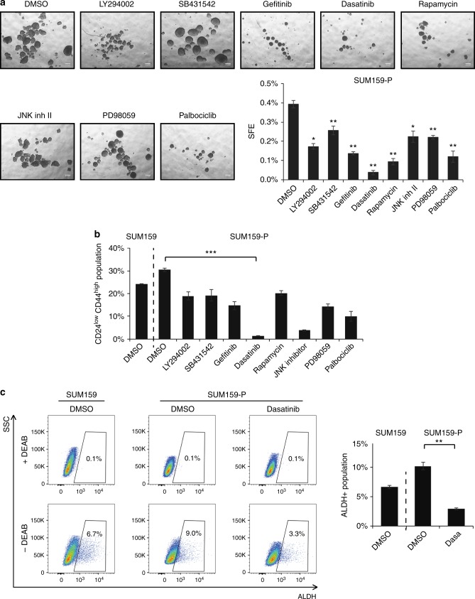
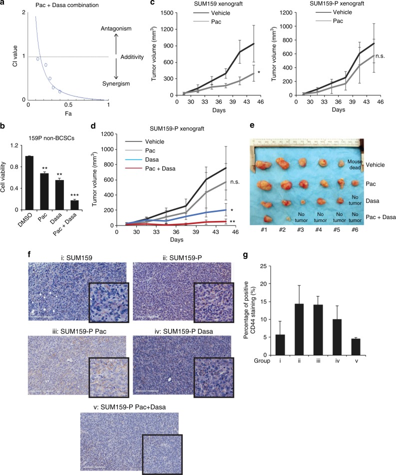
Methods: We generated paclitaxel (pac)-resistant TNBC cells which displayed higher sphere forming potential and percentage of BCSC subpopulations compared to the parental cells. A screen with various kinase inhibitors revealed dasatinib, a Src kinase family inhibitor, as a potent suppressor of BCSC expansion/sphere formation in pac-resistant TNBC cells.

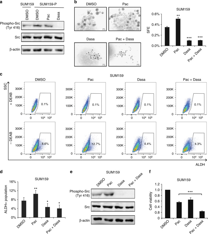
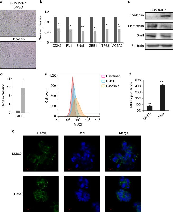
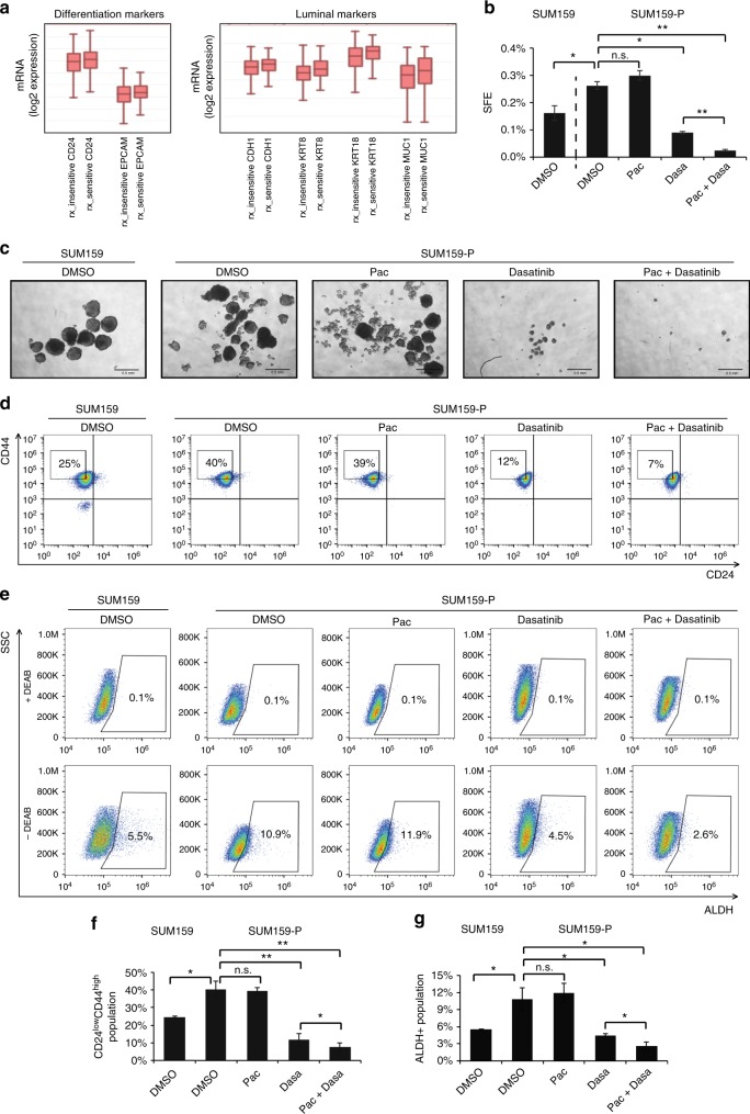
Results: We found dasatinib to block pac-induced BCSC enrichment and Src activation in both parental and pac-resistant TNBC cells. Interestingly, dasatinib induced an epithelial differentiation of the pac-resistant mesenchymal cells, resulting in their enhanced sensitivity to paclitaxel. The combination treatment of dasatinib and paclitaxel not only decreased the BCSCs numbers and their sphere forming capacity but also synergistically reduced cell viability of pac-resistant cells. Preclinical models of breast cancer further demonstrated the efficiency of the dasatinib/paclitaxel combination treatment in inhibiting tumour growth.

Conclusions: Dasatinib is a promising anti-BCSC drug that could be used in combination with paclitaxel to overcome chemoresistance in TNBC.


Conflict of interest statement

The authors declare no competing interests.

Figures

Fig. 1
Fig. 1
Paclitaxel resistance is related to stem-like properties. a Cell viability inhibition by different doses of paclitaxel in SUM159 and paclitaxel-resistant SUM159 cells (SUM159-P). The IC50 values of paclitaxel after 48 h of treatment were determined in both cell lines. b Phase-contrast microscopic images showed cell morphology of SUM159 and SUM159-P cells. c, d mRNA expression levels of ALDH1A3, CD44, MUC1 and EPCAM in breast cancer patients from the Korde dataset from Oncomine (www.oncomine.com) (n = 21, 18, 21, at 0-, 1-, 4-cycle of docetaxel, respectively). e, f Representative images of SUM159- and SUM159-P-derived tumourspheres. The number of tumourspheres (> 60 µm diameter) was counted and sphere forming efficiency (SFE) was calculated. g, h Flow cytometry analysis of ALDH+ BCSCs in SUM159 and SUM159-P cells. DEAB, a specific ALDH inhibitor, was used as a control to determine the ALDH activity. The percentage of ALDH+ populations is graphed. i, j Flow cytometry analysis of CD24lowCD44high BCSCs in SUM159 and SUM159-P cells. CD24lowCD44high population was gated based on high 50% of CD44+ population and low 50% of CD24− population. The percentage of CD24lowCD44high populations is indicated. k, l the percentage of CD24lowCD44high population in cells dissociated from SUM159 and SUM159-P derived tumours. Data are graphed as box and whisker plot, n = 3
Fig. 2
Fig. 2
Dasatinib potently kills BCSCs in SUM159-P cells. a SUM159-P cells were treated in the presence of either DMSO or IC50 doses of kinase inhibitors and subjected to tumoursphere formation assay. The SFE was calculated and graphed. b SUM159 and SUM159-P cells treated with either DMSO or IC50 doses of kinase inhibitors were subjected to flow cytometry analysis. The percentage of CD24lowCD44high populations was indicated and graphed. c SUM159 and SUM159-P cells were treated with either DMSO or IC50 dose of dasatinib. ALDEFluor assay was performed and percentage of ALDH+ cells were determined by flow cytometry
Fig. 3
Fig. 3
Dasatinib suppresses BCSC through down-regulation of paclitaxel-induced Src kinase activation. a SUM159 cells treated with DMSO and SUM159-P cells treated with DMSO, paclitaxel (10 nM), dasatinib (1 uM) were subjected with western blot analysis using anti-phospho-Src (Tyr 416), anti-Src and anti-β-actin antibodies. b Tumourspheres were derived from SUM159 cells treated with paclitaxel, dasatinib or in combination. The number of tumourspheres and SFE were determined. c, d ALDEFluor assay was conducted in SUM159 cells treated with paclitaxel, dasatinib or in combination for 4 days. The percentage of ALDH+ cells was indicated and graphed. e, f SUM159 cells were treated with paclitaxel, dasatinib or in combination for 2 days. Total cell lysates were analysed for phospho-Src (Tyr 416), Src and β-actin by western blot (e). Cell growth inhibition was measured by PrestoBlue assay (f)
Fig. 4
Fig. 4
Dasatinib induces an epithelial- and luminal-like phenotype and differentiation of chemo-resistant cells. a Cell morphology (20× magnification) of SUM159-P cells treated with DMSO or dasatinib (1 uM) for 4 days. b, c SUM159-P cells were treated with DMSO or dasatinib for 2 days. Then the mRNA and protein levels of indicated genes were subjected to real-time qPCR and immunoblotting analysis. d MUC1 mRNA expression was analysed in SUM159-P cells treated with or without dasatinib. e, f Percentage of MUC1+ population was analysed by flow cytometry in SUM159-P cells treated with dasatinib. g SUM159-P cells were treated with or without dasatinib and grown in 3D culture (Materials and Methods). Then cells were stained with antibody to F-actin (green). Nucleus was counter stained with DAPI (blue). Scale bar = 10 µm
Fig. 5
Fig. 5
Dasatinib-induced differentiation sensitises BCSCs to chemotherapy. a mRNA expression levels of the indicated genes in breast cancer patients, Booser dataset from ‘R2: Genomics Analysis and Visualisation Platform (http://r2.amc.nl)’. bg SUM159-P cells were treated with paclitaxel (10 nM), dasatinib (1 uM) or in combination. SUM159 cells were used as a control. b, c Tumoursphere formation assay was performed and the SFE was calculated. d, f Flow cytometry analysis was conducted and the percentage of CD24lowCD44high population was assessed. e, g ALDEFluor assay was conducted and percentage of ALDH+ cells were quantified by flow cytometry
Fig. 6
Fig. 6
Combination of dasatinib and paclitaxel potently eliminated breast cancer cells. a Combination index (CI)-fraction affected (Fa, corresponding to the fraction of cell viability inhibited) plot of SUM159-P cells treated with a dose range of paclitaxel in combination with dose range of dasatinib, based on a ratio of the IC50 of each drug. b CD24+CD44+ non-BCSCs were isolated from SUM159-P cells and subjected to PrestoBlue cell viability assay after the treatment of indicated drugs. ce Mice were inoculated with SUM159 and SUM159-P cells and randomly grouped (n = 6/group). The treatment started as described in the Material and Methods section. c Analyses of tumour growth in SUM159 and SUM159-P derived tumours treated with or without paclitaxel. The primary tumour volume was calculated by the formula: volume (mm3) = (length (mm))2 × (width (mm)) × 0.5. d Tumour growth curve of SUM159-P derived tumours treated with vehicle, paclitaxel, dasatinib or in combination. e Images of SUM159-P derived tumours in each treatment group. f, g CD44 expression in tumour xenografts from different groups of mice. i: SUM159 xenograft. ii: SUM159-P xenograft treated with vehicle. iii: SUM159-P xenograft treated with paclitaxel alone. iv: SUM159-P xenograft treated with dasatinib alone. v: SUM159-P xenograft treated with paclitaxel and dasatinib. The score of staining intensity and the percentage of stained area were analysed. The total percentage of membrane and cytoplasmic CD44 staining was calculated for each tumour slide. f Representative immunohistochemistry images (20×) of CD44 in breast cancer samples. Scale bar = 200 uM. g Average percentage of CD44+ area in tumour cells for each group of mice

Similar articles

Cited by

References

    1. Schneider BP, et al. Triple-negative breast cancer: risk factors to potential targets. Clin. Cancer Res. 2008;14:8010–8018. doi: 10.1158/1078-0432.CCR-08-1208. - DOI - PubMed
    1. Andre F, Zielinski CC. Optimal strategies for the treatment of metastatic triple-negative breast cancer with currently approved agents. Ann. Oncol. 2012;23:vi46–51. - PubMed
    1. Magee JA, Piskounova E, Morrison SJ. Cancer stem cells: impact, heterogeneity, and uncertainty. Cancer Cell. 2012;21:283–296. doi: 10.1016/j.ccr.2012.03.003. - DOI - PMC - PubMed
    1. Wicha MS, Liu S, Dontu G. Cancer stem cells: an old idea—a paradigm shift. Cancer Res. 2006;66:1883–1890. doi: 10.1158/0008-5472.CAN-05-3153. - DOI - PubMed
    1. Al-Hajj M, Wicha MS, Benito-Hernandez A, Morrison SJ, Clarke MF. Prospective identification of tumorigenic breast cancer cells. Proc. Natl Acad. Sci. USA. 2003;100:3983–3988. doi: 10.1073/pnas.0530291100. - DOI - PMC - PubMed

Publication types

MeSH terms

Grants and funding