Sympathetic Hyperactivity and Age Affect Segregation and Expression of Neurotransmitters
- PMID: 30483061
- PMCID: PMC6243098
- DOI: 10.3389/fncel.2018.00411
Sympathetic Hyperactivity and Age Affect Segregation and Expression of Neurotransmitters
Abstract
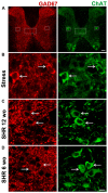
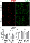
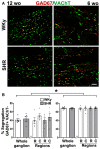
Sympathetic neurons of the rat superior cervical ganglion (SCG) can segregate their neurotransmitters and co-transmitters to separate varicosities of single axons. We have shown that transmitter segregation is a plastic phenomenon and that it is correlated with the strength of synaptic transmission. Here, we determined whether sympathetic dysfunction occurring in stress and hypertension was correlated with plastic changes of neurotransmitter segregation. We characterized the expression of the markers, L-glutamic acid decarboxylase of 67 kDa (GAD67) and vesicular acetylcholine (ACh) transporter (VAChT) in the SCG of cold stressed and spontaneously hypertensive rats (SHR). Considering that the SCG comprises a heterogeneous neuronal population, we explored whether the expression and segregation of neurotransmitters would also have an intraganglionic heterogeneous distribution in ganglia of stressed and hypertensive rats. Furthermore, since hypertension in SHR is detected around 8-10 weeks, we evaluated expression and segregation of ACh and GABA in adult hypertensive (12-week old (wo)) and young pre-hypertensive (6-wo) SHR. We found an increase in segregation of ACh and GABA with no change in transmitter expression in ganglia of stressed animals. In contrast, in SHR, there was an increase in GABA expression, although segregation did not vary. Segregation showed a caudo-rostral gradient in controls but not in the ganglia of stressed animals. GABA expression showed a rostro-caudal gradient in adult SHR, which was not present in young 6-wo rats. In young SHR, ACh increased and, unexpectedly, segregation of ACh and GABA was higher than in adults. Data suggest that ACh and GABA segregation increases in acute sympathetic hyperactivity like stress, but does not vary in chronic hyperactivity such as in hypertension. Changes in segregation are age-dependent and might be involved in the mechanisms underlying stress and hypertension.
Keywords: GABA; SHR; acetylcholine; co-transmission; hypertension; stress; sympathetic ganglia.
Figures


Similar articles
-
Functional Implications of Neurotransmitter Segregation.Front Neural Circuits. 2021 Oct 13;15:738516. doi: 10.3389/fncir.2021.738516. eCollection 2021. Front Neural Circuits. 2021. PMID: 34720888 Free PMC article. Review.
-
Ganglionic Long-Term Potentiation in Prehypertensive and Hypertensive Stages of Spontaneously Hypertensive Rats Depends on GABA Modulation.Neural Plast. 2019 Oct 13;2019:7437894. doi: 10.1155/2019/7437894. eCollection 2019. Neural Plast. 2019. PMID: 31737063 Free PMC article.
-
Age-Dependent Changes in the Occurrence and Segregation of GABA and Acetylcholine in the Rat Superior Cervical Ganglia.Int J Mol Sci. 2024 Feb 23;25(5):2588. doi: 10.3390/ijms25052588. Int J Mol Sci. 2024. PMID: 38473838 Free PMC article.
-
Segregation of Acetylcholine and GABA in the Rat Superior Cervical Ganglia: Functional Correlation.Front Cell Neurosci. 2016 Apr 7;10:91. doi: 10.3389/fncel.2016.00091. eCollection 2016. Front Cell Neurosci. 2016. PMID: 27092054 Free PMC article.
-
Hypernoradrenergic innervation: its relationship to functional and hyperplastic changes in the vasculature of the spontaneously hypertensive rat.Blood Vessels. 1989;26(1):1-20. Blood Vessels. 1989. PMID: 2540863 Review.
Cited by
-
Functional Implications of Neurotransmitter Segregation.Front Neural Circuits. 2021 Oct 13;15:738516. doi: 10.3389/fncir.2021.738516. eCollection 2021. Front Neural Circuits. 2021. PMID: 34720888 Free PMC article. Review.
-
Ganglionic Long-Term Potentiation in Prehypertensive and Hypertensive Stages of Spontaneously Hypertensive Rats Depends on GABA Modulation.Neural Plast. 2019 Oct 13;2019:7437894. doi: 10.1155/2019/7437894. eCollection 2019. Neural Plast. 2019. PMID: 31737063 Free PMC article.
-
Age-Dependent Changes in the Occurrence and Segregation of GABA and Acetylcholine in the Rat Superior Cervical Ganglia.Int J Mol Sci. 2024 Feb 23;25(5):2588. doi: 10.3390/ijms25052588. Int J Mol Sci. 2024. PMID: 38473838 Free PMC article.
References
-
- Burnstock G. (1990). The fifth heymans memorial lecture-ghent, February 17, 1990. Co-transmission. Arch. Int. Pharmacodyn. Ther. 304, 7–33. - PubMed
-
- Chan-Palay V., Palay S. L. (1984). Coexistence of Neuroactive Substances in Neurons. New York, NY: John Wiley & Sons.
LinkOut - more resources
Full Text Sources

