. 2018 Nov 13:12:85.
doi: 10.3389/fnana.2018.00085. eCollection 2018.

Innervation of the Nose and Nasal Region of the Rat: Implications for Initiating the Mammalian Diving Response

Affiliations

Innervation of the Nose and Nasal Region of the Rat: Implications for Initiating the Mammalian Diving Response

Paul F McCulloch et al. Front Neuroanat. .

Abstract

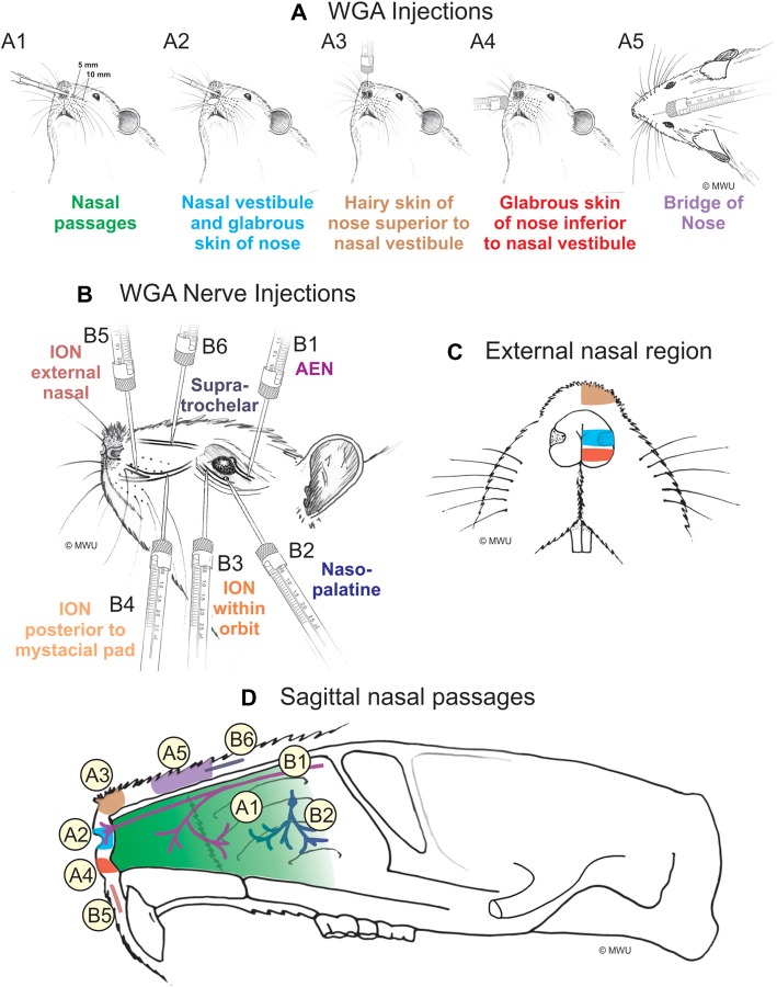
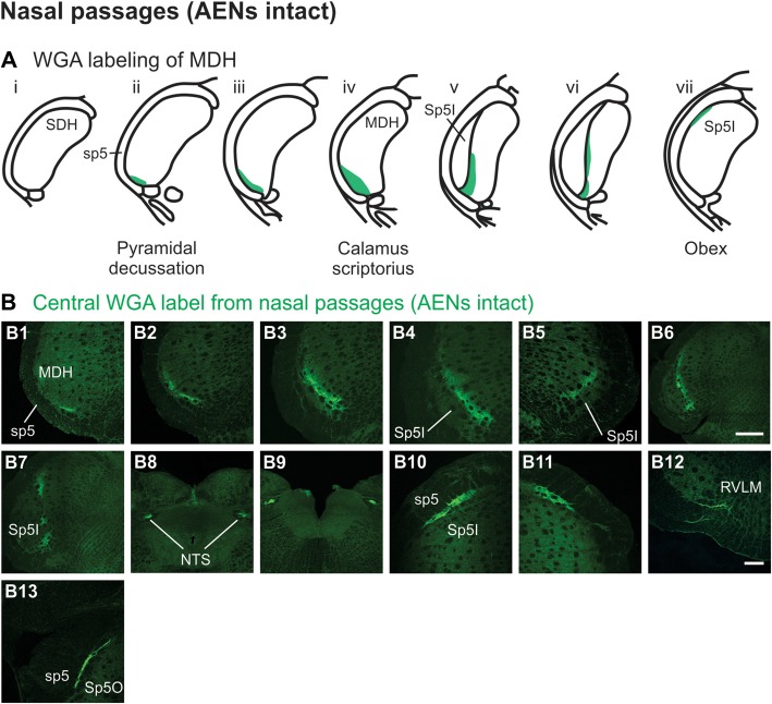
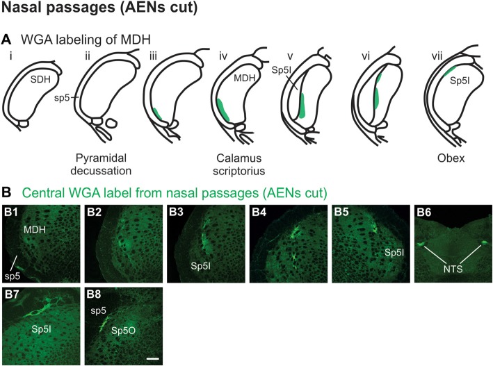
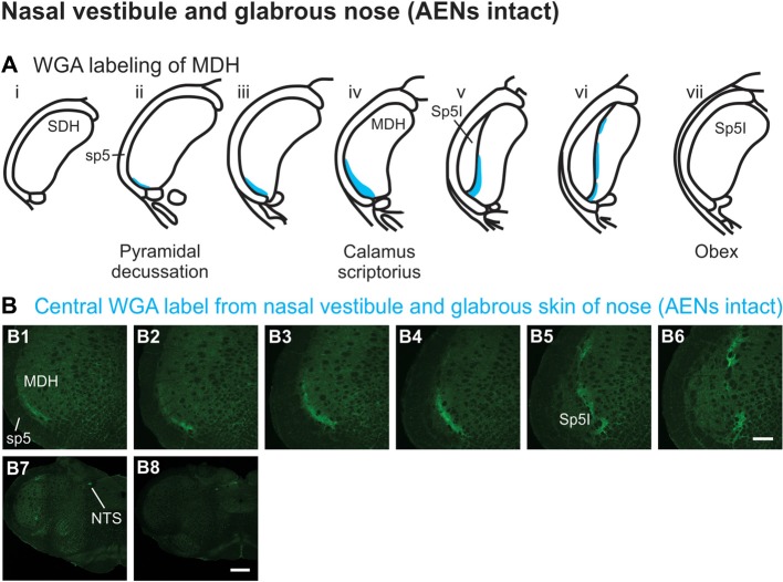
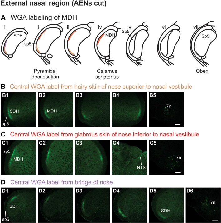
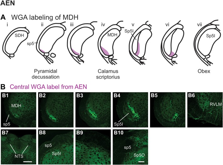
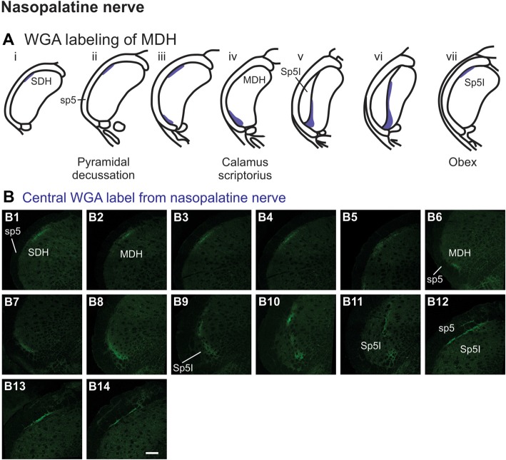
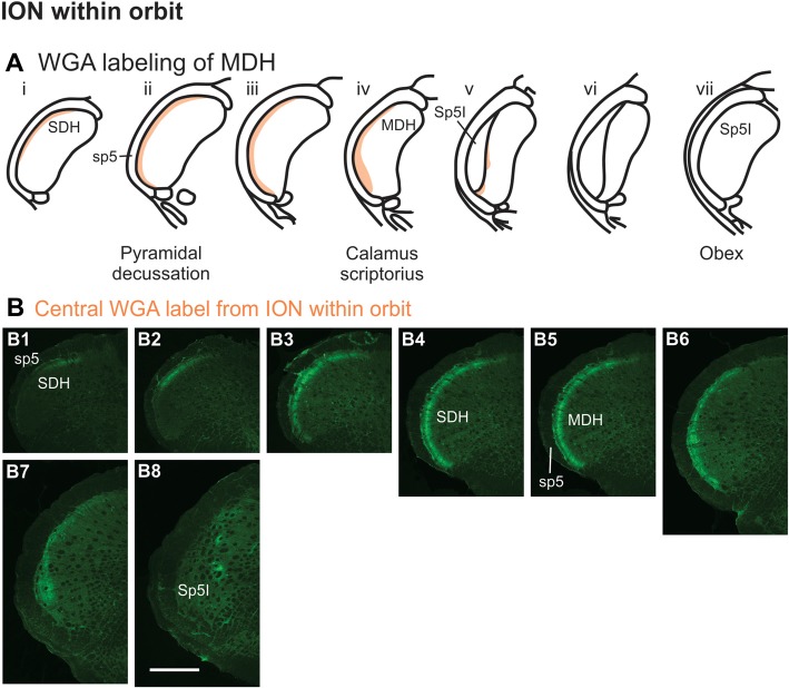
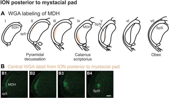
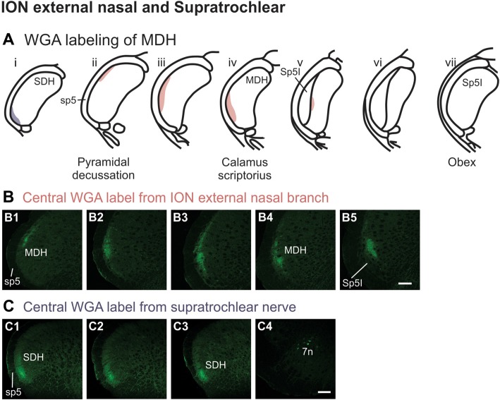
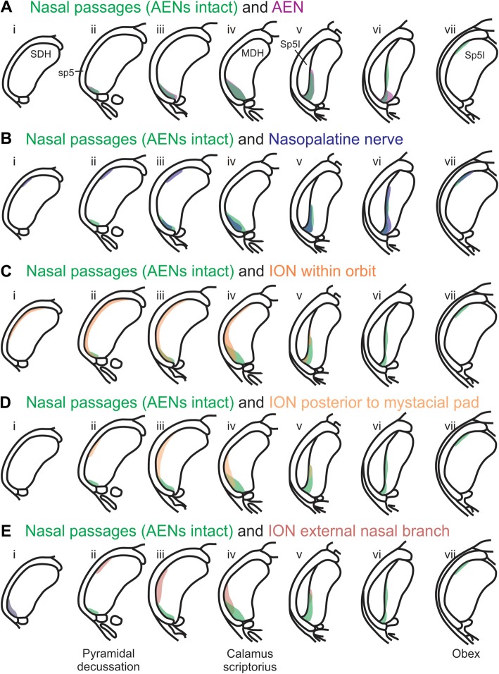
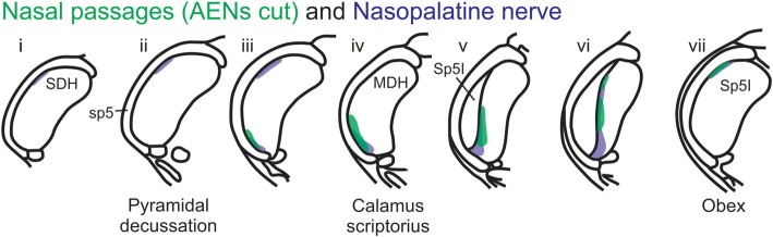
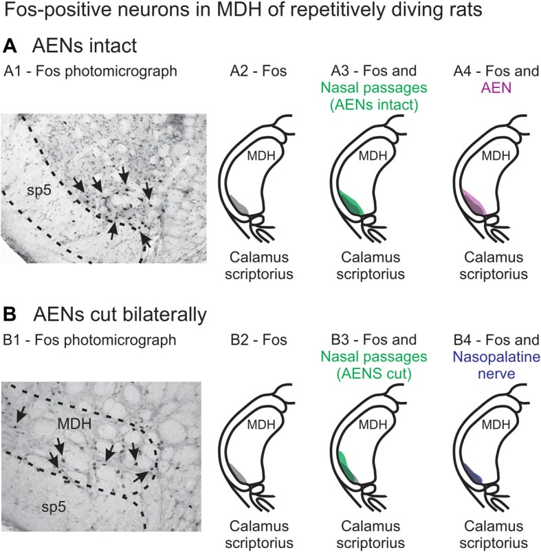
Most terrestrial animals demonstrate an autonomic reflex that facilitates survival during prolonged submersion under water. This diving response is characterized by bradycardia, apnea and selective increases in peripheral vascular resistance. Stimulation of the nose and nasal passages is thought to be primarily responsible for providing the sensory afferent signals initiating this protective reflex. Consequently, the primary objective of this research was to determine the central terminal projections of nerves innervating the external nose, nasal vestibule and nasal passages of rats. We injected wheat germ agglutinin (WGA) into specific external nasal locations, into the internal nasal passages of rats both with and without intact anterior ethmoidal nerves (AENs), and directly into trigeminal nerves innervating the nose and nasal region. The central terminations of these projections within the medulla were then precisely mapped. Results indicate that the internal nasal branch of the AEN and the nasopalatine nerve, but not the infraorbital nerve (ION), provide primary innervation of the internal nasal passages. The results also suggest afferent fibers from the internal nasal passages, but not external nasal region, project to the medullary dorsal horn (MDH) in an appropriate anatomical way to cause the activation of secondary neurons within the ventral MDH that express Fos protein during diving. We conclude that innervation of the anterior nasal passages by the AEN and nasopalatine nerve is likely to provide the afferent information responsible for the activation of secondary neurons within MDH during voluntary diving in rats.

Keywords: afferent pathways; anterior ethmoidal nerve; diving response; nasal cavity; nasopalatine nerve; nose; spinal trigeminal nucleus; trigeminal nerve.


Figures

Figure 1
Figure 1
Wheat germ agglutinin (WGA) injection sites into left nose and nasal regions. (A) Diagram showing locations of WGA injections in and around nasal region: (A1) 5 and 10 mm deep into the nasal passages; (A2) immediately inside the left nasal vestibule and brushed on the glabrous skin of the left nostril immediately distal to the nasal vestibule; (A3) into the hairy skin of the nose immediately superior to the left nasal vestibule; (A4) into the glabrous skin of the nose immediately inferior to the left nasal vestibule; and (A5) under skin along the bridge of the nose midway between the nose and the left eye. (B) Diagram showing locations of WGA injections directly into nerves: (B1) into the left anterior ethmoidal nerve (AEN) within the orbit; (B2) into the left nasopalatine nerve as it exited the orbit through the sphenopalatine foramen; (B3) into the left infraorbital nerve (ION) as it traversed the infraorbital groove deep within the orbit; (B4) into the left ION immediately posterior to the mystacial pad; (B5) into the left external nasal nerve immediately distal to the nasal vestibule, and (B6) into the left supratrochlear nerve midway between the nose tip and the left eye. (C) Summary of WGA injection sites into external nasal regions. Color-coded identification of WGA injection sites follows that from panel (A). (D) Summary of WGA injection sites in a sagittal view of internal nasal passages and external nasal region. Color-coded identification of WGA injection sites follows that from panels (A,B).
Figure 2
Figure 2
WGA label from nasal passages after WGA was injected into the left nasal passages with AENs intact (see Figure 1A1). (A) Summary cross-sections showing WGA labeling of medullary dorsal horn (MDH). (B) Photomicrographs showing central labeling within the brainstem. Label was found caudally in the ventral tip of the ipsilateral superficial MDH (B1–B3). As Sp5I began to appear ventrolaterally, label within MDH was found more medially both ipsilaterally (B4,B6,B7) and contralaterally (B5). Label was also found within NTS (B8,B9). Rostral to obex label was found bilaterally along the dorsolateral edge of Sp5I (B10,B11). Just caudal to the facial nucleus, label extended from the ventral tip of the trigeminal tract into the rostral ventrolateral medulla (B12). More rostrally, label was found as a thin strip in the dorsolateral trigeminal tract adjacent to spinal trigeminal nucleus oralis (B13). Rostral-caudal orientation: pyramidal decussation: caudal to (B1); calamus scriptorius: (B3); obex: rostral to (B7,B9). Scale bar in (B6) for (B6–B9) = 500 μm. Scale bar in (B12) for all other panels = 200 μm.
Figure 3
Figure 3
WGA label from nasal passages after WGA was injected into the left nasal passages with AENs cut bilaterally (see Figure 1A1). (A) Summary cross-sections showing WGA labeling of MDH. (B) Photomicrographs showing central labeling within the brainstem. Label was found caudally within the ipsilateral superficial MDH (B1,B2). As Sp5I appeared ventrolaterally, labeling of the dorsal MDH was found both ipsilaterally (B3,B4) and contralaterally (B5). Label was also found within NTS (B6). Rostral to obex label was found ipsilaterally along the dorsolateral edge of Sp5I (B7). More rostrally, label was found bilaterally as a thin strip in the dorsolateral trigeminal tract adjacent to the spinal trigeminal nucleus oralis (B8). Rostral-caudal orientation: calamus scriptorius: rostral to (B1); obex: rostral to (B4,B5). Scale bar in (B8) = 200 μm.
Figure 4
Figure 4
WGA label from nasal vestibule after WGA introduced immediately inside the left nasal vestibule and brushed on the glabrous skin of the left nostril immediately distal to the nasal vestibule with AENs intact (see Figure 1A2). (A) Summary cross-sections showing WGA labeling of MDH. (B) Photomicrographs showing central labeling within the brainstem. Label was found caudally in the ventral tip of ipsilateral superficial MDH (B1–B3). As Sp5I appeared ventrolaterally, label within MDH was more medial (B4–B7). Label was also found within NTS (B7). When tracer was injected into these same locations in animals with bilaterally sectioned AENs, no label was found within the ventral MDH but was found within NTS (B8). Rostral-caudal orientation: pyramidal decussation: caudal to (B1); calamus scriptorius: (B4) and (B8); obex: immediately rostral to (B7). Scale bar in (B8) for (B7) and (B8) = 500 μm. Scale bar in (B6) for all other panels = 200 μm.
Figure 5
Figure 5
WGA label from external nasal region with AENs cut bilaterally. (A) Summary cross-sections showing WGA labeling of MDH. (B) Photomicrographs showing central labeling within the brainstem after WGA was injected into the hairy skin of the nose immediately superior to the left nasal vestibule (see Figure 1A3). Label was found within the ventral tip of the superficial laminae of the spinal and medullary dorsal horn both ipsilaterally (B1–B3) and contralaterally (B4). Label was found within the ipsilateral 7n (B5). Rostral-caudal orientation: pyramidal decussation: (B2). Scale bar in (B5) for all panels = 200 μm. (C) Photomicrographs showing central labeling within the brainstem after WGA was injected into the glabrous skin of the nose immediately inferior to the left nasal vestibule (see Figure 1A4). Label was found within the superficial laminae of the lateral and dorsolateral MDH both ipsilaterally (C1,C2) and contralaterally (C3). Label was found along the edge of the ipsilateral dorsolateral NTS (C4), and ipsilateral 7n (C5). Rostral-caudal orientation: pyramidal decussation: caudal to (C1); calamus scriptorius: (C2). Scale bar in (C5) for all panels = 200 μm. (D) Photomicrographs showing central labeling within the brainstem after WGA was injected under the hairy skin along the bridge of the nose, left of the midline between the nose and left eye (see Figure 1A5). Label was found within the superficial laminae of the dorsolateral SDH (D1), lateral SDH (D2,D3), and ventrolateral tip of SDH (D4,D5). Label was found within the ipsilateral 7n (D6). Rostral-caudal orientation: pyramidal decussation: rostral to (D5). Scale bar in (D6) for all panels = 200 μm.
Figure 6
Figure 6
WGA label from AEN (see Figure 1B). (A) Summary cross-sections showing WGA labeling of MDH. (B) Photomicrographs showing central labeling within the brainstem after WGA was injected into the left AEN. Label was found caudally in the ventral tip of the ipsilateral superficial MDH (B1–B3). As Sp5I appeared ventrolaterally, label within the MDH was more medial (B4,B5). Label extended from the ventral tip of the trigeminal tract into the rostral ventrolateral medulla (B6). No label was found within NTS (B7), along the dorsolateral edge of Sp5I adjacent to the trigeminal tract (B8,B9), or in the trigeminal tract adjacent to the dorsolateral spinal trigeminal nucleus oralis (B10). Rostral-caudal orientation: pyramidal decussation: caudal to (B1); calamus scriptorius: (B3); obex: rostral to (B5,B8,B9). Scale bar in (B7) = 500 μm. Scale bar in (B10) for all other panels = 200 μm.
Figure 7
Figure 7
WGA label from nasopalatine nerve (see Figure 1B2). (A) Summary cross-sections showing WGA labeling of MDH. (B) Photomicrographs showing central labeling within the brainstem after WGA was injected into the left nasopalatine nerve. Three different patterns of labeling were found. The first pattern was found in the caudal ipsilateral dorsolateral superficial MDH (B1–B5). The second pattern was found in the ipsilateral ventrolateral superficial MDH (B6–B11). Label was found caudally in the ventrolateral tip of superficial MDH (B6–B8). As Sp5I appeared ventrolaterally label within MDH was more medial (B9–B11). The third pattern was found ipsilaterally along the dorsolateral edge of Sp5I (B12–B14). Rostral-caudal orientation: for panels (B1–B5)—pyramidal decussation: (B2); for panels (B6–B11)—calamus scriptorius: (B7); for panels (B12–B14)—obex: (B12). Scale bar in (B14) for all panels = 200 μm.
Figure 8
Figure 8
WGA label from orbital ION (see Figure 1B3). (A) Summary cross-sections showing WGA labeling of MDH. (B) Photomicrographs showing central labeling within the brainstem after WGA was injected into individual branches of the left ION as they passed through the infraorbital groove deep within the orbit. Caudally, within the spinal cord, label was first found more dorsally (B1,B2), but then more laterally (B3,B4). Label was then found throughout the entire dorsal to ventral extreme of the ipsilateral superficial MDH (B5–B7). As Sp5I appeared ventrolaterally, label within MDH was found more medially and dorsally (B8). Rostral-caudal orientation: pyramidal decussation: (B5); calamus scriptorius: (B7); obex: rostral to (B8). Scale bar in (B8) for all panels = 500 μm.
Figure 9
Figure 9
WGA label from ION immediately posterior to the mystacial pad (Figure 1B4). (A) Summary cross-sections showing WGA labeling of MDH. (B) Photomicrographs showing central labeling within the brainstem after WGA was injected into a branch of the left ION containing both external nasal and superior labial nerves immediately posterior to the mystacial pad. Label was found laterally in the caudal ipsilateral superficial MDH (B1,B2). Label was then found more ventrolaterally (B3). As Sp5I appeared ventrolaterally, label within MDH was found more medially (B4). Rostral-caudal orientation: pyramidal decussation: (B1); calamus scriptorius: (B4); Scale bar in (B4) for all panels = 200 μm.
Figure 10
Figure 10
WGA label from external nasal branch of ION and supratrochlear nerve. (A) Summary cross-sections showing WGA labeling of MDH. (B) Photomicrographs showing central labeling within the brainstem after WGA was injected directly into external nasal branch of the left ION (see Figure 1B5). Caudally label was found dorsolaterally in the ipsilateral superficial MDH (B1). Label then become more lateral (B2,B3) and ventrolateral (B4). As Sp5I appeared ventrolaterally, label within MDH was found more medially (B5). Rostral-caudal orientation: pyramidal decussation: caudal to (B1); calamus scriptorius: (B4). Scale bar in (B5) for all panels = 200 μm. (C) Photomicrographs showing central labeling within the brainstem after WGA was injected into the left supratroclear nerve (see Figure 1B6). Label was found in ventrolateral tip of SDH, approximately 1000 μm caudal to the pyramidal decussation (C1–C3). Label was found within the ipsilateral 7n (C4). Rostral-caudal orientation: pyramidal decussation: approximately 900 μm rostral to (C3). Scale bar in (C4) for all panels = 200 μm.
Figure 11
Figure 11
Summary of MDH labeling after WGA injection into the left nasal passages, along with WGA injections into nerves that innervate the left nasal region. Labeling pattern from the nasal passages with the AENs intact (from Figure 2) is shown in transparency overlays with labeling patterns from individual nerves: (A) WGA into AEN (from Figure 6); (B) WGA into nasopalatine nerve (from Figure 7); (C) WGA injection into main ION bundle as it traversed the infraorbital groove deep within the orbit with AENs intact (from Figure 8); (D) WGA injection into a branch of ION immediately posterior to the mystacial pad (from Figure 9); and (E) WGA injection into external nasal branch of ION (from Figure 10). See Figure 1 for nerve orientation and color coding of WGA injections.
Figure 12
Figure 12
Summary of MDH labeling after WGA injection into the left nasal passages and WGA injection into nasopalatine nerve. Labeling pattern from the nasal passages with the AENs cut bilaterally (from Figure 3) is shown in transparency overlay with the labeling pattern from nasopalatine nerve (from Figure 7).
Figure 13
Figure 13
Fos-positive neurons in MDH of repetitively diving rats from (A) rats with intact AENs and (B) rats with AENs cut bilaterally. Photomicrographs in (A1) from McCulloch (2005) and (B1) from McCulloch et al. (2016) show brainstem tissue immunohistologically stained to identify Fos-positive neurons from repetitively diving rats. Rostral-caudal level is just rostral to calamus scriptorius. Arrows point to Fos-positive neurons in MDH. Cross-hatching in (A2) and (B2) indicate regions of Fos-positive neurons from photomicrographs in (A1) and (B1), respectively. Area of Fos labeling (A2) is shown in transparency overlay with (A3) the WGA labeling pattern from the nasal passages with the AENs intact (from Figure 2Aiv) and with (A4) the WGA labeling pattern from the AEN (from Figure 6Aiv). Area of Fos labeling (B2) is shown in transparency overlay with (B3) the WGA labeling pattern from the nasal passages with the AENs cut bilaterally (from Figure 3Aiv) and with (B4) the WGA labeling pattern from the nasopalatine nerve (from Figure 7Aiv). Photomicrographs from McCulloch (2005) and McCulloch et al. (2016) are used with permission.

Similar articles

Cited by

References

    1. Altschuler S. M., Bao X., Bieger D., Hopkins D. A., Miselis R. R. (1989). Viscerotropic representation of the upper alimentary tract in the rat: sensory ganglia and nuclei of the solitary and spinal trigeminal tracts. J. Comp. Neurol. 283, 248–268. 10.1002/cne.902830207 - DOI - PubMed
    1. Andersson J. P., Liner M. H., Fredsted A., Schagatay E. K. (2004). Cardiovascular and respiratory responses to apneas with and without face immersion in exercising humans. J. Appl. Physiol. 96, 1005–1010. 10.1152/japplphysiol.01057.2002 - DOI - PubMed
    1. Anton F., Peppel P. (1991). Central projections of trigeminal primary afferents innervating the nasal mucosa: a horseradish peroxidase study in the rat. Neuroscience 41, 617–628. 10.1016/0306-4522(91)90354-q - DOI - PubMed
    1. Arvidsson J. (1982). Somatotopic organization of vibrissae afferents in the trigeminal sensorynuclei of the rat studied by transganglionic transport of HRP. J. Comp. Neurol. 211, 84–92. 10.1002/cne.902110108 - DOI - PubMed
    1. Arvidsson J., Rice F. (1991). Central projections of primary sensory neurons innervating different parts of the vibrissae follicles and intervibrissal skin on the mystacial pad of the rat. J. Comp. Neurol. 309, 1–16. 10.1002/cne.903090102 - DOI - PubMed