

Distinct Role of IL-27 in Immature and LPS-Induced Mature Dendritic Cell-Mediated Development of CD4+ CD127+3G11+ Regulatory T Cell Subset
- PMID: 30483251
- PMCID: PMC6244609
- DOI: 10.3389/fimmu.2018.02562
Distinct Role of IL-27 in Immature and LPS-Induced Mature Dendritic Cell-Mediated Development of CD4+ CD127+3G11+ Regulatory T Cell Subset
Abstract
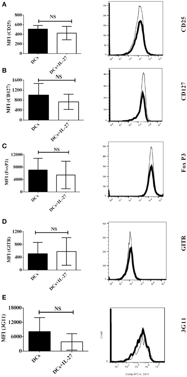
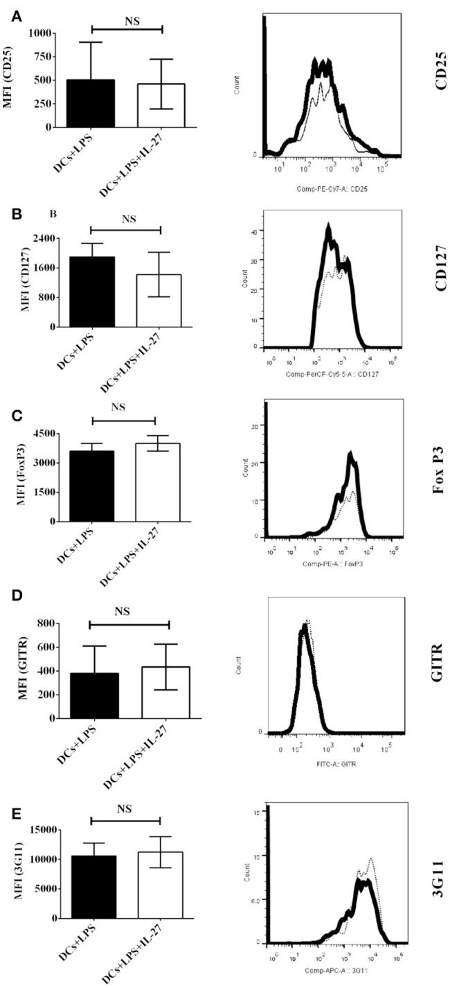
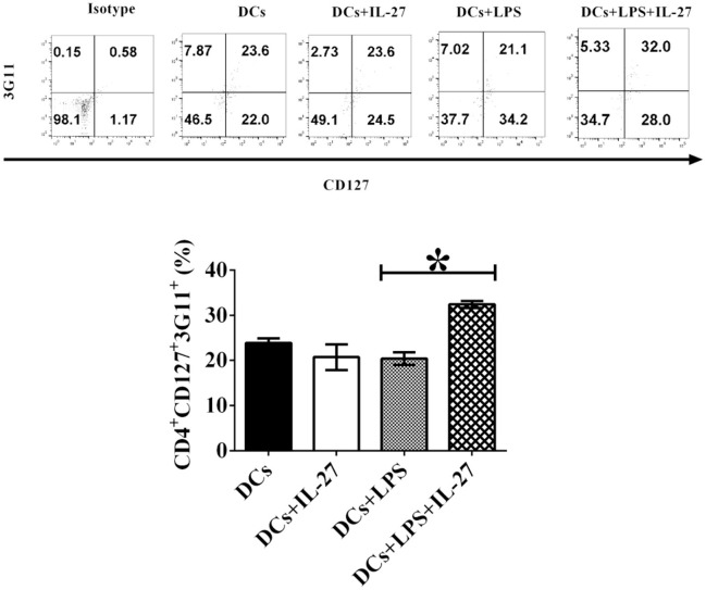
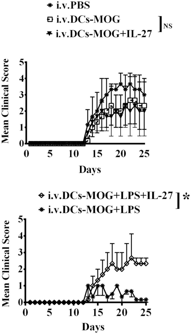
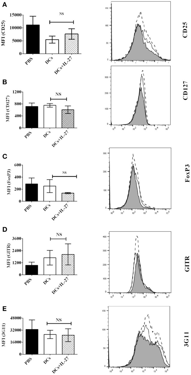
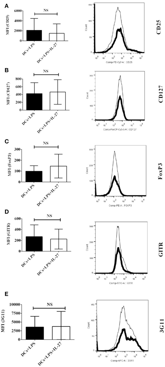
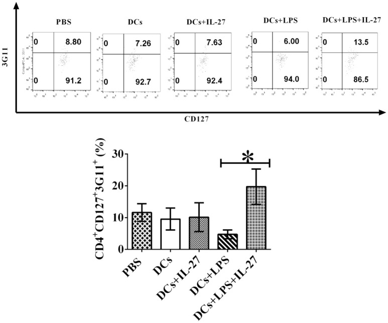
Interleukin-27 (IL-27) plays an important role in regulation of anti-inflammatory responses and autoimmunity; however, the molecular mechanisms of IL-27 in modulation of immune tolerance and autoimmunity have not been fully elucidated. Dendritic cells (DCs) play a central role in regulating immune responses mediated by innate and adaptive immune systems, but regulatory mechanisms of DCs in CD4+ T cell-mediated immune responses have not yet been elucidated. Here we show that IL-27 treated mature DCs induced by LPS inhibit immune tolerance mediated by LPS-stimulated DCs. IL-27 treatment facilitates development of the CD4+ CD127+3G11+ regulatory T cell subset in vitro and in vivo. By contrast, IL-27 treated immature DCs fail to modulate development of the CD4+CD127+3G11+ regulatory T cell sub-population in vitro and in vivo. Our results suggest that IL-27 may break immune tolerance induced by LPS-stimulated mature DCs through modulating development of a specific CD4+ regulatory T cell subset mediated by 3G11 and CD127. Our data reveal a new cellular regulatory mechanism of IL-27 that targets DC-mediated immune responses in autoimmune diseases such as multiple sclerosis (MS) and experimental autoimmune encephalomyelitis (EAE).
Keywords: IL-27; dendritic cell; immune tolerance; immunotherapy; regulatory T cell.
Figures
Similar articles
-
LPS-treated bone marrow-derived dendritic cells induce immune tolerance through modulating differentiation of CD4+ regulatory T cell subpopulations mediated by 3G11 and CD127.Immunol Res. 2017 Jun;65(3):630-638. doi: 10.1007/s12026-016-8881-z. Immunol Res. 2017. PMID: 27942984 Free PMC article.
-
Immune tolerance induced by intravenous transfer of immature dendritic cells via up-regulating numbers of suppressive IL-10(+) IFN-γ(+)-producing CD4(+) T cells.Immunol Res. 2013 May;56(1):1-8. doi: 10.1007/s12026-012-8382-7. Immunol Res. 2013. PMID: 23292714 Free PMC article.
-
1,25-dihydroxyvitamin D3 -induced dendritic cells suppress experimental autoimmune encephalomyelitis by increasing proportions of the regulatory lymphocytes and reducing T helper type 1 and type 17 cells.Immunology. 2017 Nov;152(3):414-424. doi: 10.1111/imm.12776. Epub 2017 Jul 10. Immunology. 2017. PMID: 28617989 Free PMC article.
-
Regulatory dendritic cells in autoimmunity: A comprehensive review.J Autoimmun. 2015 Sep;63:1-12. doi: 10.1016/j.jaut.2015.07.011. Epub 2015 Aug 5. J Autoimmun. 2015. PMID: 26255250 Review.
-
3G11 expression in CD4+ T cell-mediated autoimmunity and immune tolerance.Int Immunopharmacol. 2011 May;11(5):593-6. doi: 10.1016/j.intimp.2010.11.005. Epub 2010 Nov 24. Int Immunopharmacol. 2011. PMID: 21084064 Free PMC article. Review.
Cited by
-
Crosstalk between dendritic cells and regulatory T cells: Protective effect and therapeutic potential in multiple sclerosis.Front Immunol. 2022 Sep 13;13:970508. doi: 10.3389/fimmu.2022.970508. eCollection 2022. Front Immunol. 2022. PMID: 36177043 Free PMC article. Review.
-
Role of innate immune cells in multiple sclerosis.Front Immunol. 2025 Feb 17;16:1540263. doi: 10.3389/fimmu.2025.1540263. eCollection 2025. Front Immunol. 2025. PMID: 40034690 Free PMC article. Review.
-
Biologic Agents in Crohn's Patients Reduce CD4+ T Cells Activation and Are Inversely Related to Treg Cells.Can J Gastroenterol Hepatol. 2022 Jul 31;2022:1307159. doi: 10.1155/2022/1307159. eCollection 2022. Can J Gastroenterol Hepatol. 2022. PMID: 35959163 Free PMC article.
-
Anti-JMH alloantibody in inherited JMH-negative patients leads to immunogenic destruction of JMH-positive RBCs.Clin Exp Immunol. 2021 Aug;205(2):182-197. doi: 10.1111/cei.13622. Epub 2021 Jun 13. Clin Exp Immunol. 2021. PMID: 34021913 Free PMC article.
-
IL-27 Improves Prophylactic Protection Provided by a Dead Tumor Cell Vaccine in a Mouse Melanoma Model.Front Immunol. 2022 Apr 21;13:884827. doi: 10.3389/fimmu.2022.884827. eCollection 2022. Front Immunol. 2022. PMID: 35529885 Free PMC article.
References
MeSH terms
Substances
Grants and funding
LinkOut - more resources
Full Text Sources
Research Materials