Review
. 2018 Nov 12:9:538.
doi: 10.3389/fgene.2018.00538. eCollection 2018.

Function and Regulation of Human Terminal Uridylyltransferases

Affiliations
Review

Function and Regulation of Human Terminal Uridylyltransferases

Yuka Yashiro et al. Front Genet. .

Abstract

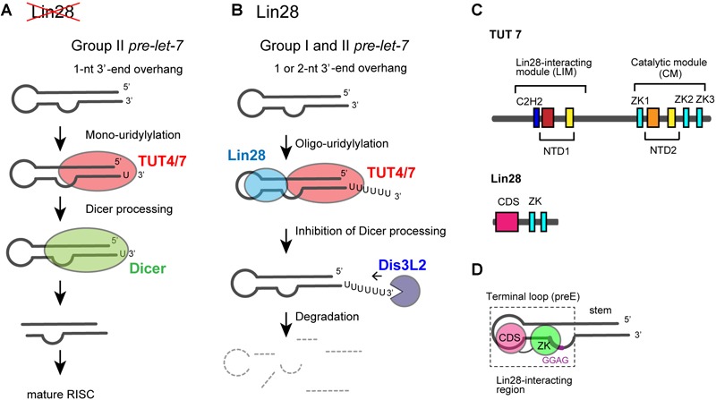
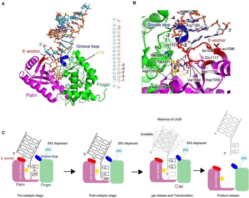
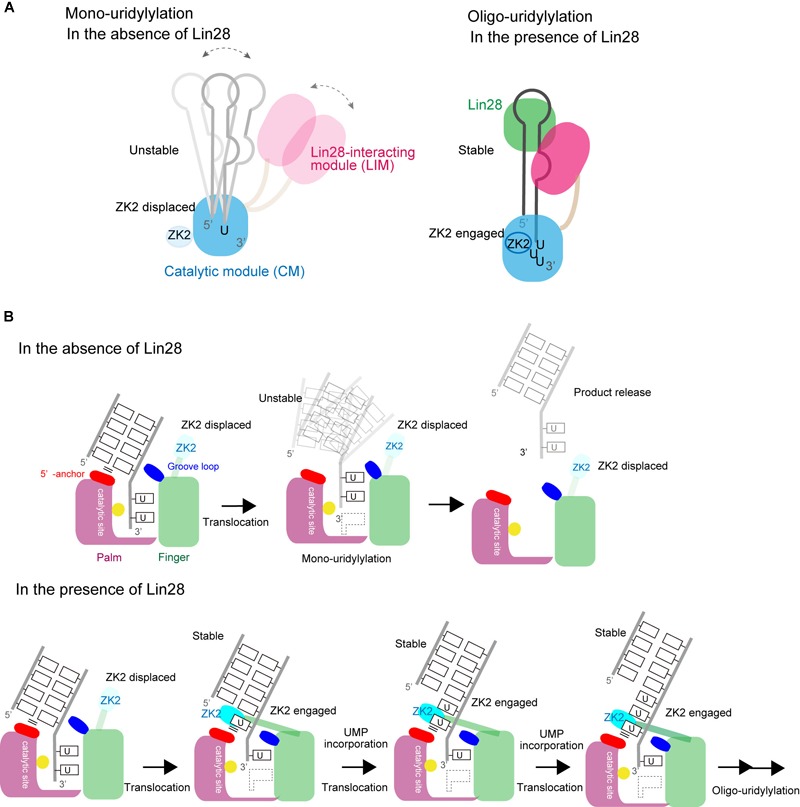
RNA uridylylation plays a pivotal role in the biogenesis and metabolism of functional RNAs, and regulates cellular gene expression. RNA uridylylation is catalyzed by a subset of proteins from the non-canonical terminal nucleotidyltransferase family. In human, three proteins (TUT1, TUT4, and TUT7) have been shown to exhibit template-independent uridylylation activity at 3'-end of specific RNAs. TUT1 catalyzes oligo-uridylylation of U6 small nuclear (sn) RNA, which catalyzes mRNA splicing. Oligo-uridylylation of U6 snRNA is required for U6 snRNA maturation, U4/U6-di-snRNP formation, and U6 snRNA recycling during mRNA splicing. TUT4 and TUT7 catalyze mono- or oligo-uridylylation of precursor let-7 (pre-let-7). Let-7 RNA is broadly expressed in somatic cells and regulates cellular proliferation and differentiation. Mono-uridylylation of pre-let-7 by TUT4/7 promotes subsequent Dicer processing to up-regulate let-7 biogenesis. Oligo-uridylylation of pre-let-7 by TUT4/7 is dependent on an RNA-binding protein, Lin28. Oligo-uridylylated pre-let-7 is less responsive to processing by Dicer and degraded by an exonuclease DIS3L2. As a result, let-7 expression is repressed. Uridylylation of pre-let-7 depends on the context of the 3'-region of pre-let-7 and cell type. In this review, we focus on the 3' uridylylation of U6 snRNA and pre-let-7, and describe the current understanding of mechanism of activity and regulation of human TUT1 and TUT4/7, based on their crystal structures that have been recently solved.

Keywords: TUT1; TUT4/7; TUTase; U6 snRNA; biogenesis; let-7; splicing; terminal uridylyltransferase.


Figures

FIGURE 1
FIGURE 1
Human non-canonical terminal nucleotidyltransferases. Schematic representation of domain organization of seven human non-canonical terminal nucleotidyltransferases. The catalytic motif is composed of nucleotidyltransferase domain (orange box) and PAP-associated domain (yellow box). Inactive nucleotidyltransferase domains are designated by red boxes. C2H2-type zinc finger and CCHC zinc finger domains are designated as dark blue and light blue boxes, respectively. RNA recognition motif (RRM), is shown as a green box. The figure is modified from Heo et al. (2009) and Lee et al. (2014).
FIGURE 2
FIGURE 2
TUT1 in the maturation process of human U6 snRNA. (A) Secondary structure of human mature U6 snRNA transcript. Mature U6 snRNA harbors 5′-γ-methyl tri-phosphate (5′-pmpp) and 2′,3′-cyclic phosphate ( > p) at 5′- and 3′-ends, respectively. (B) Maturation of U6 snRNA. Primary U6 snRNA transcript harbors four genome-encoded 3′-uridines (UUUUOH). The 3′-end is oligo-uridylylated by TUT1, with the addition of up to 20 uridines. Finally, oligo-uridylylated U6 snRNA is trimmed by Usb1. Mature U6 snRNA harbors five 3′-uridines capped with a 2′,3′-cyclic phosphate (UUUUU > p).
FIGURE 3
FIGURE 3
Structure of human TUT1. (A) The overall structure of human TUT1 lacking N-terminal ZF and RRM (TUT1_delN). Palm (magenta), fingers (green) and KA-1 (cyan). UTP (stick model in yellow) resides in the cleft between palm and finger domains. Inset, the overall structure of human TUT1 lacking C-terminal KA-1. The linker between RRM (orange) and palm domain is flexible. (B) KA-1 domain of TUT1 (upper, cyan) is homologous to KA-1 domain of MARK3 kinase (lower, pink). (C) Superposition of TUT1_delN structures of different forms of crystals. C-terminal KA-1 (cyan) domains of TUT1 are mobile and can rotate approximately 40 degrees relative to the catalytic domain, using α14 as the rotation axis. (D) Nucleotide recognition by human TUT1. UTP recognition (left) and ATP recognition (right). Nucleotides are depicted by stick models.
FIGURE 4
FIGURE 4
Interactions between U6 snRNA and TUT1. (A) Electrostatic potential of KA-1 domain of human TUT1. Positively and negatively charged areas are colored blue and red, respectively. KA-1 domain is outlined by yellow line (upper), and harbors clusters of positively charged amino acids (below). (B) Multi-domain utilization by TUT1 for U6 snRNA oligo-uridylylation. Schematic representation of interactions between U6 snRNA and TUT1 analyzed by Tb(III) hydrolysis mapping. N-terminal ZF and RRM (orange), catalytic palm and fingers (green), and C-terminal KA-1 (cyan). (C). Mechanism of oligo-uridylylation of U6 snRNA by TUT1. KA-1 and ZF-RRM are mobile. Binding of TUT1 to U6 snRNA induces conformational change of U6 snRNA, and KA-1 of TUT1 acts as an anchor during oligo-uridylylation. N-terminal ZF and RRM (orange), catalytic palm and fingers (green), and C-terminal KA-1 (cyan).
FIGURE 5
FIGURE 5
Functional duality of TUT4/7 in the biogenesis of let-7. (A) In the absence of LIN28, mono-uridylylation of pre–let-7 that harbors 1-nt 3′-overhang (group II) by TUT4/7 promotes Dicer processing of pre–let-7. (B) In the presence of LIN28, TUT4/7 oligo-uridylylates pre-let-7 and inhibits Dicer processing of pre–let-7. Oligo-uridylylated pre–let-7 is degraded by DIS3L2, an exonuclease. (C) Schematic representation of domain organization of TUT4/7 and Lin28. ZKs in Lin28 interact with LIM of TUT4/7 in the presence of pre–let-7 (Faehnle et al., 2017; Wang et al., 2017). (D) Schematic representation of the secondary structure of pre–let-7 and interactions with Lin28B (Nam et al., 2011; Wang et al., 2017). ZK of Lin28 binds GGAG motif in pre–let-7 and CDS binds the terminal loop of preE.
FIGURE 6
FIGURE 6
Structure of human TUT7 representing mono-uridylylation. (A) The overall structure of TUT7 CM complexed with dsRNA that mimics the double-helix of group II pre–let-7 and UTP. ZK2 was not visible in the structure suggesting that the ZK2 is displaced. (B) UTP recognition by TUT7. UTP is depicted by sticks. (C) Schematic representation of mono-uridylylation of pre–let-7 with 1-nt 3′-overhang (group II). After mono-uridylylation of group II pre–let-7 and pyrophosphate (ppi) release, pre–let-7 tanslocates. In the absence of Lin28, the mono-uridylylated pre–let-7 cannot form a stable complex with TUT7, and is easily released from TUT7.
FIGURE 7
FIGURE 7
Structure of human TUT7 reflecting oligo-uridylylation. (A) Structure of TUT7 CM complexed with UUOH and UTP, representing the pre-catalytic stage. (B) Structure of TUT7 CM complexed with UUUUUOH (U5), representing the post-catalytic stage. (C) Schematic representation of oligo-uridylylation of pre–let-7 with 2-nt 3′-overhang (group II). ZK2 participates in the recognition of uridine at position -2 to stabilize the 3′-oligo(U) in the pre- (left) and post- (right) catalytic stages.
FIGURE 8
FIGURE 8
Switching between mono- and oligo-uridylylation. (A) Schematic representation of mono-uridylylation in the absence of Lin28 (left) and oligo-uridylylation in the presence of Lin28 (right). (B) Schematic detailed representations of mono-uridylylation of pre–let-7 in the absence of Lin28 (upper), and oligo-uridylylation of pre–let-7 in the presence of Lin28 (lower). Only the catalytic site in the CM is presented.

Similar articles

Cited by

References

    1. Achsel T., Brahms H., Kastner B., Bachi A., Wilm M., Luhrmann R. (1999). A doughnut-shaped heteromer of human Sm-like proteins binds to the 3’-end of U6 snRNA, thereby facilitating U4/U6 duplex formation in vitro. EMBO J. 18 5789–5802. 10.1093/emboj/18.20.5789 - DOI - PMC - PubMed
    1. Aphasizhev R., Suematsu T., Zhang L., Aphasizheva I. (2016). Constructive edge of uridylation-induced RNA degradation. RNA Biol. 13 1078–1083. 10.1080/15476286.2016.1229736 - DOI - PMC - PubMed
    1. Aravind L., Koonin E. V. (1999). DNA polymerase beta-like nucleotidyltransferase superfamily: identification of three new families, classification and evolutionary history. Nucleic Acids Res. 27 1609–1618. 10.1093/nar/27.7.1609 - DOI - PMC - PubMed
    1. Astuti D., Morris M. R., Cooper W. N., Staals R. H., Wake N. C., Fews G. A., et al. (2012). Germline mutations in DIS3L2 cause the Perlman syndrome of overgrowth and Wilms tumor susceptibility. Nat. Genet. 44 277–284. 10.1038/ng.1071 - DOI - PubMed
    1. Bai Y., Srivastava S. K., Chang J. H., Manley J. L., Tong L. (2011). Structural basis for dimerization and activity of human PAPD1, a noncanonical poly(A) polymerase. Mol. Cell 41 311–320. 10.1016/j.molcel.2011.01.013 - DOI - PMC - PubMed