

Induction of Apoptosis in Toxoplasma gondii Infected Hela Cells by Cisplatin and Sodium Azide and Isolation of Apoptotic Bodies and Potential Use for Vaccination against Toxoplasma gondii
- PMID: 30483332
- PMCID: PMC6243158
Induction of Apoptosis in Toxoplasma gondii Infected Hela Cells by Cisplatin and Sodium Azide and Isolation of Apoptotic Bodies and Potential Use for Vaccination against Toxoplasma gondii
Abstract
Background: Toxoplasma gondii can infect a wide range of mammalians, especially humans. It controls several intracellular signals for the inhibition of apoptosis. This study aimed to investigate the apoptogenic effect of cisplatin and sodium azide on T. gondii infected HeLa cells and isolate apoptotic bodies (blebs) as a potent stimulator of the immune system.
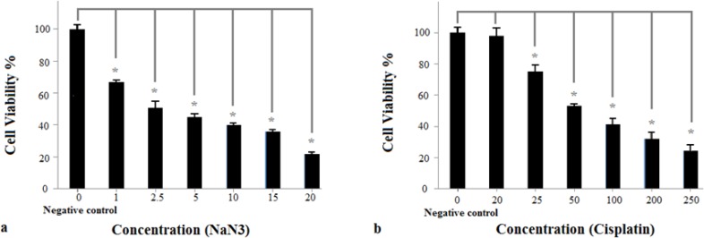
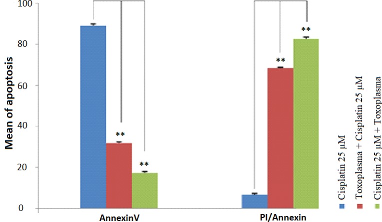
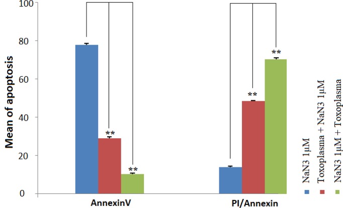
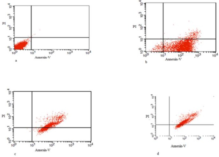
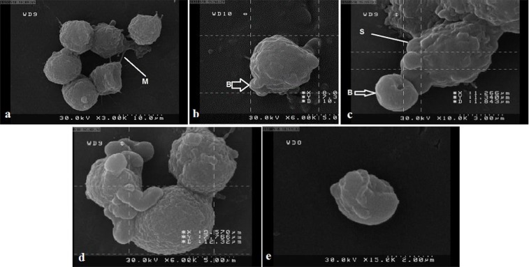
Methods: The cytotoxic properties of cisplatin and sodium azide (NaN3) on HeLa cells were evaluated by MTT assay. Moreover, the apoptogenic activity of cisplatin and NaN3 was studied using flow cytometry (Annexin V/PI double staining) and scanning electron microscopy (SEM). Finally, apoptotic bodies were separated by centrifugation.
Results: MTT assay data showed that the survival rate of cells treated with different concentration of NaN3 was significantly reduced, compared to negative control groups. Concerning cisplatin, only concentration of 20 μM had not a significant impact on the cell viability; however, the other concentration of cisplatin significantly reduced cell viability, compared to negative control groups. The level of early apoptosis in uninfected HeLa cells was higher compared to infected HeLa cells treated with cisplatin and NaN3. Finally, apoptotic bodies were separated from T. gondii infected HeLa cells treated with cisplatin.
Conclusion: Apoptosis was induced in both uninfected and infected HeLa cells with T. gondii and apoptotic bodies were isolated from infected cells. Therefore, further studies on apoptotic bodies are required in order to find a proper candidate for vaccine preparation against T. gondii infections.
Keywords: Apoptosis; Apoptotic blebs; Cisplatin; NaN3; Toxoplasma gondii; Toxoplasmosis.
Conflict of interest statement
Conflict of interest The authors declare that there is no conflict of interests.
Figures
Similar articles
-
Toxoplasma gondii infection confers resistance against BimS-induced apoptosis by preventing the activation and mitochondrial targeting of pro-apoptotic Bax.J Cell Sci. 2009 Oct 1;122(Pt 19):3511-21. doi: 10.1242/jcs.050963. Epub 2009 Sep 8. J Cell Sci. 2009. PMID: 19737817
-
Biogenic silver nanoparticles (AgNp-Bio) reduce Toxoplasma gondii infection and proliferation in HeLa cells, and induce autophagy and death of tachyzoites by apoptosis-like mechanism.Acta Trop. 2021 Oct;222:106070. doi: 10.1016/j.actatropica.2021.106070. Epub 2021 Jul 28. Acta Trop. 2021. PMID: 34331897
-
Toxoplasma gondii infection inhibits the mitochondrial apoptosis through induction of Bcl-2 and HSP70.Parasitol Res. 2010 Nov;107(6):1313-21. doi: 10.1007/s00436-010-1999-3. Epub 2010 Aug 3. Parasitol Res. 2010. PMID: 20680337
-
Toxoplasma gondii Modulates the Host Cell Responses: An Overview of Apoptosis Pathways.Biomed Res Int. 2019 Apr 4;2019:6152489. doi: 10.1155/2019/6152489. eCollection 2019. Biomed Res Int. 2019. PMID: 31080827 Free PMC article. Review.
-
Apoptosis and its modulation during infection with Toxoplasma gondii: molecular mechanisms and role in pathogenesis.Curr Top Microbiol Immunol. 2005;289:219-37. doi: 10.1007/3-540-27320-4_10. Curr Top Microbiol Immunol. 2005. PMID: 15791958 Review.
Cited by
-
Tailoring of apoptotic bodies for diagnostic and therapeutic applications:advances, challenges, and prospects.J Transl Med. 2024 Sep 1;22(1):810. doi: 10.1186/s12967-024-05451-w. J Transl Med. 2024. PMID: 39218900 Free PMC article. Review.
References
-
- Mohaghegh MA, Yazdani H, Hadipour M, et al. Seroprevalence of Toxoplasma gondii infection among patient admitted to Al-zahra hospital, Isfahan, IRAN. J Ayub Med Coll Abbottabad. 2015;27:767–70. - PubMed
-
- Derouin F, Pelloux H. Prevention of toxoplasmosis in transplant patients. Clin Microbiol Infect. 2008;14:1089–101. - PubMed
-
- Awan KJ. Congenital toxoplasmosis: chances of occurrence in subsequent siblings. Ann Ophthalmol. 1978;10:459–65. - PubMed
-
- Kodjikian L, Hoigne I, Adam O, et al. Vertical transmission of toxoplasmosis from a chronically infected immunocompetent woman. Pediatr Infect Dis J. 2004;23:272–4. - PubMed
-
- Vogel N, Kirisits M, Michael E, et al. Congenital toxoplasmosis transmitted from an immunologically competent mother infected before conception. Clin Infect Dis. 1996;23:1055–60. - PubMed
LinkOut - more resources
Full Text Sources
Other Literature Sources
Research Materials
Miscellaneous