

The effects of bortezomib on the ovariectomy applied rat uterus: A histopathological, stereological, and immunohistochemical study
- PMID: 30483384
- PMCID: PMC6251398
- DOI: 10.22038/IJBMS.2018.24756.6152
The effects of bortezomib on the ovariectomy applied rat uterus: A histopathological, stereological, and immunohistochemical study
Abstract
Objectives: In this study, potential protective effects of Bortezomib (Bort), as a proteasome inhibitor, were investigated on the uterus of ovariectomized rats by histological, morphometric and immunohistochemical methods.
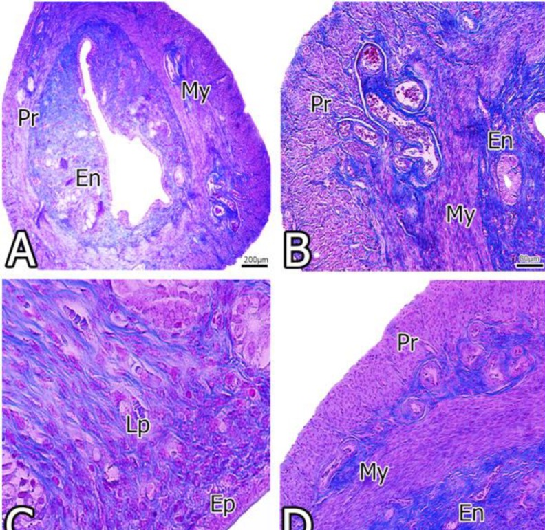
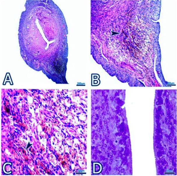
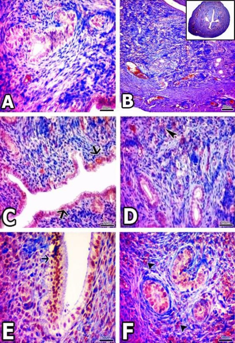
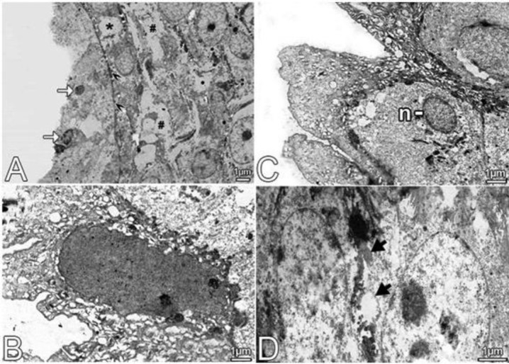
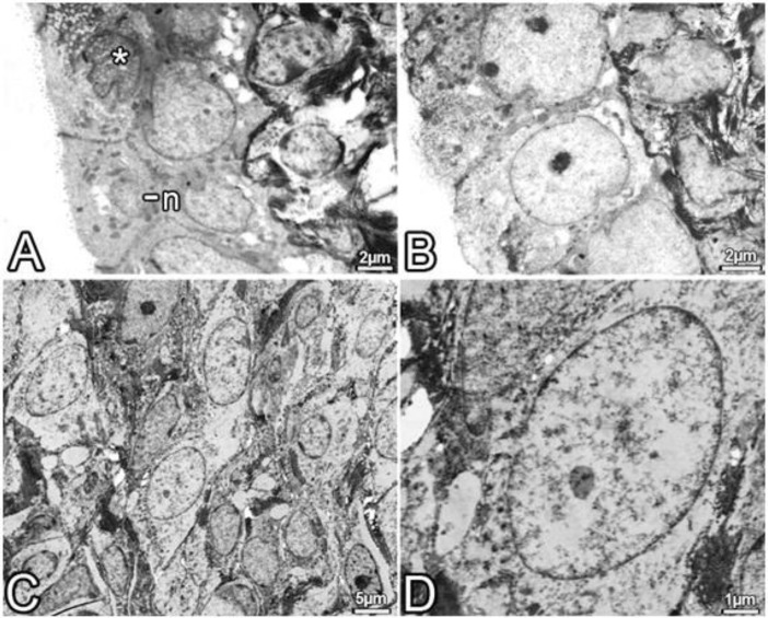
Materials and methods: In this study, 18 Sprague dawley strain female rats (12 weeks old, 250-300 g body weight) were used. Animals in the control group (Cont, n=6) were not exposed to any treatment. Ovariectomy was performed on the experimental groups. They (n=12) were divided into ovariectomy (Ovt, n=6) and Bortezomib (Bort, n=6) subgroups. Twelve weeks later, the rats were perfused. Then, uterine tissues were removed and examined by morphometrical, and light and electron microscopy methods. In addition, immunoreactivity of nuclear factor-kappa (NF-κB) was evaluated.
Results: Morphometric and histopathological evaluations showed that Bort was effective in the uterus and protects the layer structures and the cells.
Conclusion: In the light of these findings, we suggest that for proteasome inhibitor particularly Bort is thought to be useful through proteasome inhibition and NF-κB pathway.
Keywords: Bortezomib; Microscopy; Ovariectomy; Rat; Stereology; Uterus.
Conflict of interest statement
The authors declare that they have no conflicts of interest.
Figures


Similar articles
-
The protective effect of the proteasome inhibitor bortezomib on the uterus of ovariectomized rats.Clin Exp Obstet Gynecol. 2016;43(5):737-741. Clin Exp Obstet Gynecol. 2016. PMID: 30074329
-
Antiosteoporotic activity of echinacoside in ovariectomized rats.Phytomedicine. 2013 Apr 15;20(6):549-57. doi: 10.1016/j.phymed.2013.01.001. Epub 2013 Feb 18. Phytomedicine. 2013. PMID: 23428402
-
Bortezomib activation of mTORC1 pathway mediated by NOX2-drived reactive oxygen species results in apoptosis in primary dorsal root ganglion neurons.Exp Cell Res. 2021 Mar 15;400(2):112494. doi: 10.1016/j.yexcr.2021.112494. Epub 2021 Jan 27. Exp Cell Res. 2021. PMID: 33515593
-
Characterization of the pharmacologic profile of a standardized soy extract in the ovariectomized rat model of menopause: effects on bone, uterus, and lipid profile.Menopause. 2005 Sep-Oct;12(5):589-600. doi: 10.1097/01.GME.0000156348.61767.D5. Epub 2005 Sep 1. Menopause. 2005. PMID: 16145313
-
Anti-inflammatory effect of the proteasome inhibitor bortezomib on endotoxin-induced uveitis in rats.Invest Ophthalmol Vis Sci. 2012 Jun 26;53(7):3682-94. doi: 10.1167/iovs.12-9505. Invest Ophthalmol Vis Sci. 2012. PMID: 22538426
References
LinkOut - more resources
Full Text Sources