. 2018 Nov;21(11):1126-1132.
doi: 10.22038/IJBMS.2018.26688.6536.

Immunohistochemical characterization of pancreatic duodenal homeobox protein-1, neurogenin-3, and insulin protein expressions in islet-mesenchymal cell in vitro: a morphochronological evaluation

Affiliations

Immunohistochemical characterization of pancreatic duodenal homeobox protein-1, neurogenin-3, and insulin protein expressions in islet-mesenchymal cell in vitro: a morphochronological evaluation

Juziel K Manda et al. Iran J Basic Med Sci. 2018 Nov.
No abstract available

Keywords: Co-culture; Duct ligated pancreas; Insulin; Islet; MSC; NeuroG3; Pdx1; Transplantation.


Figures

Figure 1
Figure 1
Expression of pancreatic duodenal homeobox protein-1 and neuroginin-3 during early development of islets cultured by direct contact with or without mesenchymes. Immunocytochemistry analysis of whole mount islets cells isolated from PPDL and SOC and cultured without (A-D) for PDL and I-L for SOC, or with (E-H) MSCs for PDL and M-P for SOC. Islet cells development was evaluated by anti-Pdx1 (green) and anti-Ngn3 (red). PDX1/NGN3 (orange). Nuclei were counter-stained in blue with DAPI. ), Scale bar = 100 µm
Figure 2
Figure 2
Expression of pancreatic duodenal homeobox protein-1 and neurogenin-3 during the second week of development of islets cultured by direct contact with or without mesenchymes. Islets and mesenchymal cells (MSCs) were isolated from SOC and PPDL tissues respectively and cultured with (islets+ MSC-, islets/ MSC+) for different periods. Immunocytochemistry analysis of PPDL islets cells cultured without (A-D) or without (A-D) with (E-H) for 9 (A and E), 11 (B and F), 13 (C and G) and 15 (D and H) days of culture. I-P: Immunocytochemistry analysis of 24 hr SOC islets cells cultured without (I-L) or with (M-P) MSCs for same period of time. Islet cells development was evaluated by anti-Pdx1 (green) and anti-Ngn3 (orange/red). Nuclei were counter-stained in blue with DAPI. Bar = 100 µm
Figure 3
Figure 3
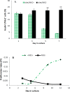
A) A bar-graph representing insulin+/pancreatic duodenal homeobox protein-1+ co-expression during the first week of development of islet/mesenchymal cell cultures of duct ligated pancreata. Quantification of absolute surface areas occupied by Insulin+Pdx1+cells that developed over 3, 5, 7 days of culture with or without mesenchyme. Forty (40) to 50 islets were analysed for each condition. Data are means±SE. **P<0.05 (P=0.0481), Scale bar =100 µm. B) A trend graph showing the proliferation index of cells in islet/mesenchymal cell cultures of duct ligated pancreata during 2 weeks of development. Forty (40) to 50 islets were analysed for each condition. Data are means ± SE. **P<0.05. Scale bar=100 µm
Figure 4
Figure 4
Representative growth in size of the islets in islet/mesenchymal cell cultures of duct ligated pancreata. Islets were grown for 28 days with or without MSCs. A and D: Islets at day 0 of culture with or without MSCs. B and E: Islets cultured for 15 days with or without MSCs. C and F: Islets cultured for 28 with or without MSCs. The surfaces of the islets after dithizone staining were marked and measured and areas of all islets were added up. G: Quantification of the total size of the islets before, after 15 and 28 days in culture in the absence or presence of MSCs. Thirty (30) to 40 islets were analysed in each condition. Data of mean values in µm2±SE are shown in the graph. **P<0.05
Figure 5
Figure 5
Expression of insulin and neurogenin-3 and the efficacy index of neurogenin-3 in islet/mesenchymal cell cultures of duct ligated pancreata. Immunocytochemistry analysis of islet cultures of PPDL tissues without (A-C) or with (D-F) MSCs. Islet cells development was evaluated by anti-Insulin (green) and anti-Ngn3 (orange/red). G: Quantification of the absolute surface areas occupied by insulin+cells and Ngn3+ cells that developed over 24, 26 or 28 days of culture with or without MSCs. Forty (40) to fifty (50) islets were analysed for each condition. Data are means±SE. *P<0.05 (P=0.037). Scale bar=100 µm

Similar articles

References

    1. Chintinne M, Stangé G, Denys B, Ling Z, Pipeleers D. Beta cell count instead of beta cell mass to assess and localize growth in beta cell population following pancreatic duct ligation in mice. PLoS One. 2012;7:e43959. - PMC - PubMed
    1. Xu X, D’Hoker J, Stange G, Bonne S, Leu N De, Xiao X, et al. β cells can be generated from endogenous progenitors in injured adult mouse pancreas. Cell. 2008;132:197–207. - PubMed
    1. Tchokonte-Nana V. Cellular mechanisms involved in the recapitulation of endocrine development in the duct ligated pancreas. Stellenbosch: University of Stellenbosch; 2011.
    1. Inada A, Nienaber C, Katsuta H, Fujitani Y, Levine J, Morita R, et al. Carbonic anhydrase II-positive pancreatic cells are progenitors for both endocrine and exocrine pancreas after birth. Proc Natl Acad Sci U S A. 2008;105:19915–19919. - PMC - PubMed
    1. Murtaugh LC, Kopinke D. Pancreatic stem cells. In: L. Charles Murtaugh, Daniel Kopinke., editors. Cambridge (MA) 2008. (StemBook) - PubMed

LinkOut - more resources