Endodontic management of central incisor associated with large periapical lesion and fused supernumerary root: a conservative approach
- PMID: 30483468
- PMCID: PMC6237732
- DOI: 10.5395/rde.2018.43.e44
Endodontic management of central incisor associated with large periapical lesion and fused supernumerary root: a conservative approach
Abstract
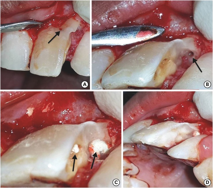
Fusion and gemination are developmental anomalies of teeth that may require endodontic treatment. Fusion may cause various clinical problems related to esthetics, tooth spacing, and other periodontal complications. Additional diagnostic tools are required for the diagnosis and the treatment planning of fused tooth. The present case report describes a case of unilateral fusion of a supernumerary root to an upper permanent central incisor with large periapical lesion in which a conservative approach was used without extraction of supernumerary tooth and obturated with mineral trioxide aggregate to reach a favorable outcome.
Keywords: Cone-beam computed tomography; Fused teeth; Mineral trioxide aggregate; Supernumerary tooth.
Conflict of interest statement
Conflict of Interest: No potential conflict of interest relevant to this article was reported.
Figures


Similar articles
-
Endodontic Management of Dilacerated Maxillary Central Incisor fused to a Supernumerary Tooth using Cone Beam Computed Tomography: An Unusual Clinical Presentation.J Contemp Dent Pract. 2017 Jun 1;18(6):522-526. doi: 10.5005/jp-journals-10024-2077. J Contemp Dent Pract. 2017. PMID: 28621286
-
Endodontic management of mandibular central incisor fused to a supernumerary tooth associated with a talon cusp: a case report.Int Endod J. 2012 Jun;45(6):590-6. doi: 10.1111/j.1365-2591.2012.02029.x. Epub 2012 Mar 6. Int Endod J. 2012. PMID: 22394311
-
Fusion of central incisors with supernumerary teeth: a 10-year follow-up of multidisciplinary treatment.J Endod. 2014 Jul;40(7):1020-4. doi: 10.1016/j.joen.2013.12.004. Epub 2014 Jan 10. J Endod. 2014. PMID: 24935556
-
Fusion of a supernumerary tooth to right mandibular second molar: a case report and literature review.Int J Clin Exp Med. 2015 Aug 15;8(8):11890-5. eCollection 2015. Int J Clin Exp Med. 2015. PMID: 26550101 Free PMC article. Review.
-
Hemisection and vital treatment of a fused tooth--literature review and case report.Endod Dent Traumatol. 1997 Dec;13(6):253-8. doi: 10.1111/j.1600-9657.1997.tb00051.x. Endod Dent Traumatol. 1997. PMID: 9558505 Review.
Cited by
-
Fusion of a Tooth with a Supernumerary Tooth: A Case Report and Literature Review of 35 Cases.Children (Basel). 2023 Dec 20;11(1):6. doi: 10.3390/children11010006. Children (Basel). 2023. PMID: 38275427 Free PMC article.
-
Fusion of Unerupted Mesiodens with a Regular Maxillary Central Incisor: a Diagnostic and Therapeutic Challenge.Acta Stomatol Croat. 2021 Sep;55(3):325-331. doi: 10.15644/asc55/3/10. Acta Stomatol Croat. 2021. PMID: 34658379 Free PMC article.
References
-
- De Jonge TE. Geminate tooth formation: schizodontia and synodontia, abstracted. Dent Abstr. 1957;2:41.
-
- Rani A K, Metgud S, Yakub SS, Pai U, Toshniwal NG, Bawaskar N. Endodontic and esthetic management of maxillary lateral incisor fused to a supernumerary tooth associated with a talon cusp by using spiral computed tomography as a diagnostic aid: a case report. J Endod. 2010;36:345–349. - PubMed
-
- Hagman FT. Anomalies of form and number, fused primary teeth, a correlation of the dentitions. ASDC J Dent Child. 1988;55:359–361. - PubMed
-
- Kayalibay H, Uzamis M, Akalin A. The treatment of a fusion between the maxillary central incisor and supernumerary tooth: report of a case. J Clin Pediatr Dent. 1996;20:237–240. - PubMed
Publication types
LinkOut - more resources
Full Text Sources