. 2018 Nov 13:8:517.
doi: 10.3389/fonc.2018.00517. eCollection 2018.

High Therapeutic Efficacy of a New Survivin LSP-Cancer Vaccine Containing CD4+ and CD8+ T-Cell Epitopes

Affiliations

High Therapeutic Efficacy of a New Survivin LSP-Cancer Vaccine Containing CD4+ and CD8+ T-Cell Epitopes

Fanny Onodi et al. Front Oncol. .

Abstract

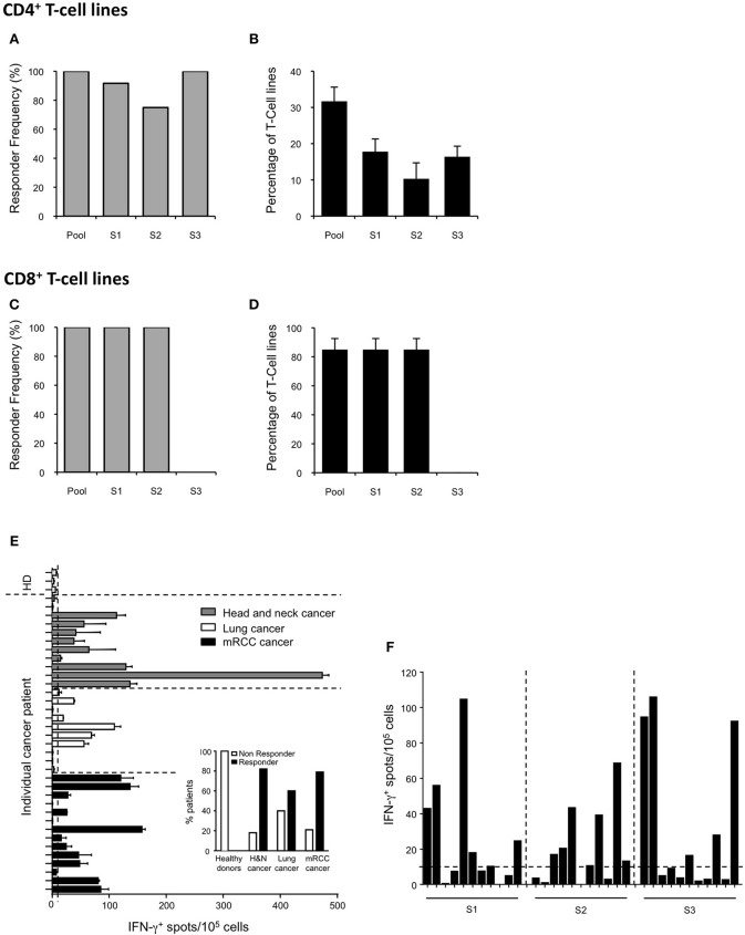
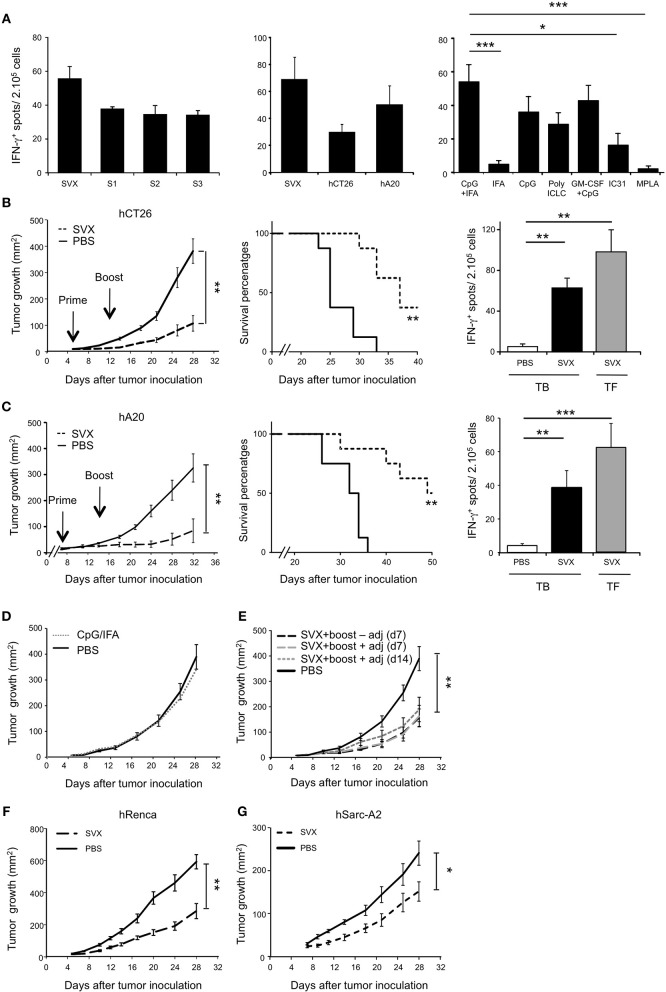
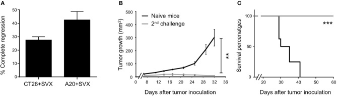
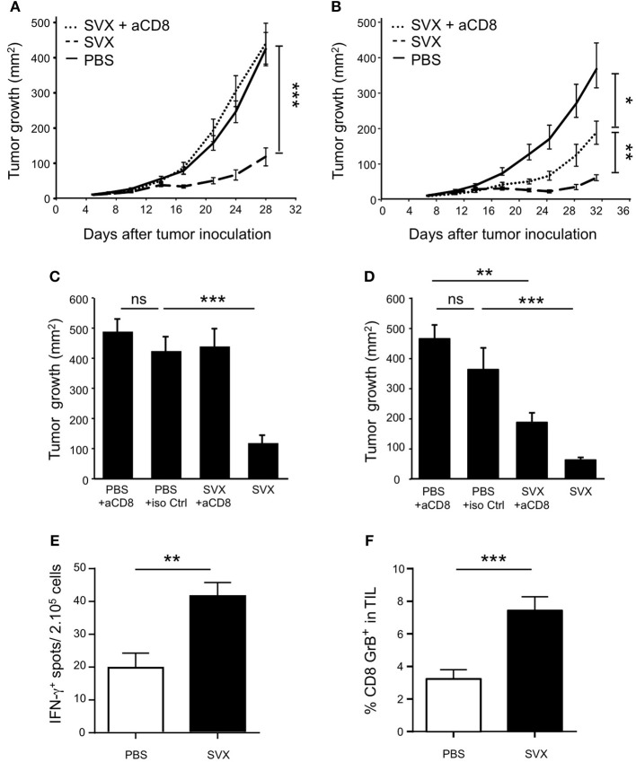
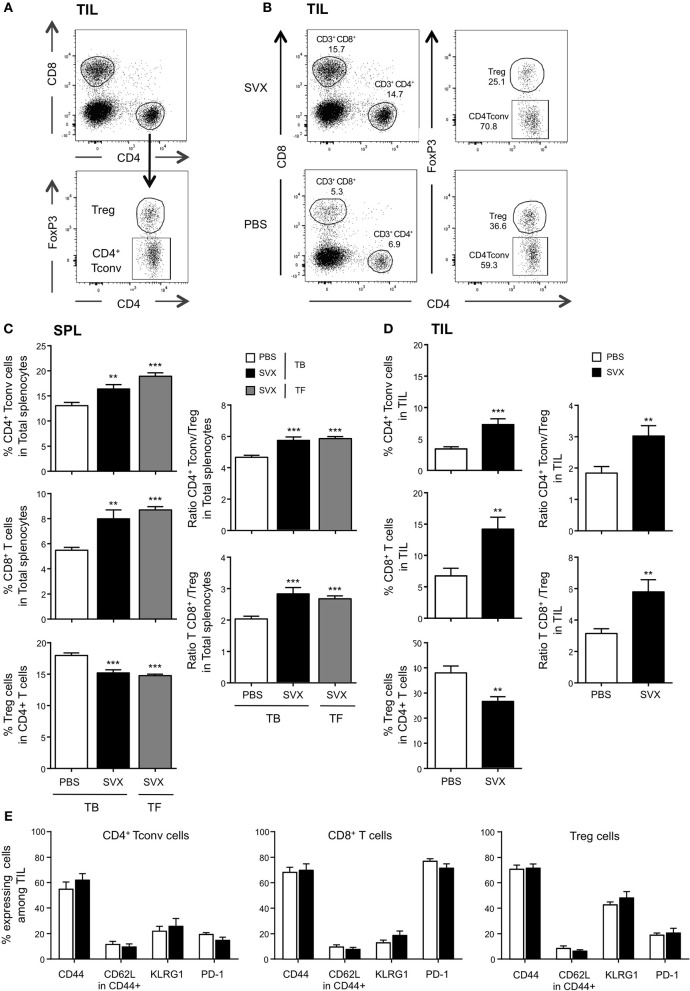
The efficacy of an antitumoral vaccine relies both on the choice of the antigen targeted and on its design. The tumor antigen survivin is an attractive target to develop therapeutic cancer vaccines because of its restricted over-expression and vital functions in most human tumors. Accordingly, several clinical trials targeting survivin in various cancer indications have been conducted. Most of them relied on short peptide-based vaccines and showed promising, but limited clinical results. In this study, we investigated the immunogenicity and therapeutic efficacy of a new long synthetic peptide (LSP)-based cancer vaccine targeting the tumor antigen survivin (SVX). This SVX vaccine is composed of three long synthetic peptides containing several CD4+ and CD8+ T-cell epitopes, which bind to various HLA class II and class I molecules. Studies in healthy individuals showed CD4+ and CD8+ T-cell immunogenicity of SVX peptides in human, irrespective of the individual's HLA types. Importantly, high frequencies of spontaneous T-cell precursors specific to SVX peptides were also detected in the blood of various cancer patients, demonstrating the absence of tolerance against these peptides. We then demonstrated SVX vaccine's high therapeutic efficacy against four different established murine tumor models, associated with its capacity to generate both specific cytotoxic CD8+ and multifunctional Th1 CD4+ T-cell responses. When tumors were eradicated, generated memory T-cell responses protected against rechallenge allowing long-term protection against relapses. Treatment with SVX vaccine was also found to reshape the tumor microenvironment by increasing the tumor infiltration of both CD4+ and CD8+ T cells but not Treg cells therefore tipping the balance toward a highly efficient immune response. These results highlight that this LSP-based SVX vaccine appears as a promising cancer vaccine and warrants its further clinical development.

Keywords: T-cell responses; cancer vaccine; immunotherapy; long synthetic peptide; tumor associated survivin antigen.


Figures

Figure 1
Figure 1
Induction of T-cell responses to SVX in healthy donors and spontaneous T-cell responses in cancer patients. (A–D) CD4+ and CD8+ T-cell responses against SVX peptides in healthy donors. CD4+ T cells from 12 healthy donors with diverse HLA-DRB1 genotype (A,B) or CD8+ T cells from 3 HLA-A*02:01 healthy donors (C,D) were repeatedly stimulated in vitro with the pool of SVX peptides (S1+S2+S3) loaded on autologous DCs. T-cell specificity was assessed by IFN-γ ELISpot assays using PBMCs or C1R-A2 loaded with the pool or individual SVX peptides. Histograms show the frequency of responding donors (A,C) and the mean percentages ± SEM of specific T-cell lines induced per donors (B,D) responding to each individual SVX peptide or at least one SVX peptide (Pool). (E,F) Spontaneous T-cell responses to SVX peptides in healthy donors, and cancer patients. (E) PBMC from 3 healthy donors, 11 head and neck, 10 lung, and 14 mRCC cancer patients were screened for spontaneous T-cell reactivity against the pool of SVX peptides, in IFN-γ ELISpot assays, after 1 week of in vitro culture with the pool of SVX peptides. (E) Intensity of the survivin response in different cancer patients and healthy donors. Each bar represents one patient or donor. Data are presented as means of IFN-γ spots from one experiment in triplicate. Small histograms represent the percentage of responder and of non-responder in healthy donors and in different types of cancer patients. (F) The 11 mRCC-responder patients were screened for spontaneous T-cell reactivity against individual SVX peptide, in IFN-γ ELISpot assays, after 1 week of in vitro culture. Data are presented as means of IFN-γ spots from one experiment in triplicate. A response was considered positive if the number of spots per well obtained in peptide(s) stimulated conditions was two-fold higher than the number of spots counted without peptide(s), with a cut-off at 10 spot-forming cells after subtracting background.
Figure 2
Figure 2
High therapeutic efficacy of SVX vaccine against various established tumor models. (A) T-cell immunogenicity of SVX vaccine and adjuvant selection. Tumor-free BALB/c (H2d) mice were subcutaneously (s.c) vaccinated as followed: priming with the three survivin LSPs (SVX) and adjuvants and boost 2 weeks later with SVX without adjuvant. One week after the boost, the induction of survivin-specific T-cell responses was analyzed by IFN-γ ELISpot assay on total splenocytes (2 × 10⁵ cells). Left and middle graphs showed overnight restimulation with the pool of SVX peptides or individual peptides (A, left graph) or tumor cell lines expressing the human survivin (hCT26 and hA20) (A, middle graph). Comparison of SVX specific T-cell responses induced by various adjuvants was performed by restimulation with the pool of SVX peptides (A, right graph). Results are the mean ± SEM of 5 mice per group and are representative of two to three independent experiments. ***P < 0.001, *P < 0.05. (B,C) Therapeutic experiments. BALB/c mice were engrafted s.c with hCT26 (2 × 10⁵ cells) (B) or hA20 cells (2.5 × 10⁵ cells) (C). When tumors reached 10 mm2, mice were s.c injected with PBS, or immunized with SVX + CpG/IFA and received a boost 1 week later without adjuvant (SVX). (B,C) (Left graphs). Tumor growth was monitored twice a week and data are presented as mean tumor size (mm2) ± SEM. **P < 0.01. (Middle graphs). Kaplan-Meier survival curves of mice treated (dashed line) or not (black line) with SVX vaccine. The experimental endpoint was applied when tumor size reached 300 mm2. (Right Graphs). Intensity of SVX specific T-cell responses in the different groups: Tumor-bearing (TB) mice injected with PBS, or vaccinated (SVX) and Tumor-free (TF) mice immunized with SVX vaccine. Data are presented as means of IFN-γ spots ± SEM in the different groups of mice. **P < 0.01, ***P < 0.001. Data are representative of the results obtained in five separate experiments with 8 mice per group. (D,E) Control studies in the hCT26 tumor model. (D) Impact of the adjuvant on tumor growth. When tumors reached 10 mm2, mice were injected with PBS (PBS), or injected with the adjuvant alone (CpG/IFA). Data represent the mean of tumor size (mm2) ± SEM. (E) Impact of the vaccination strategy at boost on the vaccine efficacy. BALB/c mice were engrafted s.c with hCT26 tumor cells and injected with PBS (PBS), or vaccinated as followed: boost at d7 without adjuvant, at d7 with SVX + adjuvant or at d14 with SVX + adjuvant. Tumor growth was monitored every 2–3 days and data are presented as mean tumor size (mm2) ± SEM. The experiment has been repeated two-three times with similar results. **P < 0.01. (F,G) BALB/c mice engrafted s.c with hRenca (5 × 10⁵ cells) (F) or humanized HLA-A2/DR1 transgenic mice engrafted s.c with hSarc-A2 (5 × 10⁵ cells) tumor cells (G) were s.c injected with PBS or vaccinated with SVX + CpG/IFA and boosted 1 week later with SVX (SVX). Data are presented as mean tumor size (mm2) ± SEM and is representative of one out of two independent experiments with 8 mice per group. *P < 0.05, **P < 0.01.
Figure 3
Figure 3
Generation of long term anti-tumor memory T cells following SVX vaccination. (A–C) Studies on tumor regression. (A) Histograms represent the percentage ± SEM of complete tumor regression of a pool of five different experiments (representing 40 mice) for both hCT26 and hA20 tumor models. (B) A group of vaccinated mice engrafted with hA20 cells (n = 5), which completely eliminated tumor cells were re-challenged with hA20 tumor cells. As positive control naive BALB/c mice (n = 5) were engrafted with hA20 tumor cells (Naive mice). Tumor growth (B) and Survival (C) were monitored. The experiment has been performed twice. **P < 0.01, ***P < 0.001.
Figure 4
Figure 4
SVX therapeutic efficacy against tumor cells in CD8-depleted mice. BALB/c mice (8 mice per group) were engrafted s.c with hCT26 (A,C) or hA20 tumor cells (B,D). When tumors reached 10 mm2, mice were s.c injected with PBS, or were immunized with SVX + CpG/IFA and received a boost 1 week later without adjuvant (SVX). (A,B) Groups of vaccinated mice engrafted with hCT26 (A) or hA20 tumor cells (B) were depleted of CD8+ T cells, using anti-CD8 mAbs (100 μg) injected intra-peritoneally (i.p) once a week, starting 1 day before SVX immunization (SVX + αCD8). Data are presented as mean tumor size (mm2) ± SEM from cohorts of 8 mice with *P < 0.05, **P < 0.01 and ***P < 0.001. Experiments have been done twice. (C,D) An additional group of mice for each experiment is shown and represents BALB/c mice (8 mice per group) engrafted with hCT26 (C) or hA20 tumors (D) and depleted of CD8+ T cells using anti-CD8 mAbs (100 μg) injected i.p once a week during 3 weeks. Data represent the mean of tumor size (mm2) ± SEM at day 28 (C) or day 32 (D). **P < 0.01 and ***P < 0.001. (E) Intensity of survivin CD8+ specific T-cell responses in hCT26 TB mice vaccinated (SVX) or not with SVX (PBS). Evaluation of functional responses was performed 2 weeks after the last vaccination using IFN-γ ELISpot assays on total splenocytes restimulated overnight in vitro with the CD8+ T-cell epitope surv85-93. Data are presented as means of IFN-γ spots ± SEM of 16 mice per group from two independent experiments. **P < 0.01. (F) Same mice as in (E). Two weeks after the last immunization, the tumors were harvested, and the percentage of CD8+GrB+ in TIL was evaluated by flow cytometry. Data are presented as means of percentage of cells ± SEM of 16 mice per group from two independent experiments. ***P < 0.001.
Figure 5
Figure 5
Cytokine profile of the CD4+ T-cell responses induced with SVX vaccine. Tumor-bearing (TB) mice engrafted s.c with hCT26 (n = 8 per group) were s.c injected with PBS or vaccinated with SVX vaccine (SVX). Tumor-free (TF) were immunized with SVX vaccine (SVX). Two weeks after the last immunization, splenic CD4+ T cells were cell sorted by magnetic beads. CD4+ T cells (2 × 10⁵) were then co-cultured with BM-DC (5 × 10⁴) pulsed with medium or the pool of SVX peptides. Cytokine productions were measured by Luminex assay performed on the supernatant after 24 h (A) or 48 h (B) of culture. Data are mean ± SEM of 8 mice per group with *P < 0.05, **P < 0.01, and ***P < 0.001.
Figure 6
Figure 6
Impact of SVX vaccine on frequencies and phenotype of T-cell subsets both among splenocytes and TIL. (A–E) Groups of mice were the same as Figure 5. Two weeks after the last immunization, the spleen (C) and the tumor (D) were harvested, the immune cells were isolated, and the percentages of CD4+ Tconv, CD8+ T cells, and CD4+ FoxP3+ Treg cells were evaluated in each individual mouse by flow cytometry. (A) Dot plots show the strategy of gating to obtain the percentages of each population. (B) Dot plots show an example of percentage of each population recovered in TIL from TB mice vaccinated or not with SVX. The expression of different cell surface markers was also assessed in the different populations of T cells isolated from the tumor (E). (C,D) (Left panels) Data are presented as means of percentage of cells ± SEM of 16 mice per group from two independent experiments. (C,D) (Right panels) The ratios of CD4+ Tconv/Treg and CD8/Treg of splenic (C) and TIL (D) were calculated for each individual mouse. Data are presented as means of ratios ± SEM of 16 mice per group from two independent experiments. **P < 0.01 and ***P < 0.001.

Similar articles

Cited by

References

    1. Kissick HT, Sanda MG. The role of active vaccination in cancer immunotherapy: lessons from clinical trials. Curr Opin Immunol. (2015) 35:15–22. 10.1016/j.coi.2015.05.004 - DOI - PubMed
    1. Karaki S, Anson M, Tran T, Giusti D, Blanc C, Oudard S, et al. . Is there still room for cancer vaccines at the Era of checkpoint inhibitors. Vaccines (2016) 4:E37. 10.3390/vaccines4040037 - DOI - PMC - PubMed
    1. van der Burg SH, Arens R, Ossendorp F, van Hall T, Melief CJM. Vaccines for established cancer: overcoming the challenges posed by immune evasion. Nat Rev Cancer (2016) 16:219–33. 10.1038/nrc.2016.16 - DOI - PubMed
    1. Speiser DE, Romero P. Molecularly defined vaccines for cancer immunotherapy, and protective T cell immunity. Semin Immunol. (2010) 22:144–54. 10.1016/j.smim.2010.03.004 - DOI - PubMed
    1. Garrido F, Algarra I. MHC antigens and tumor escape from immune surveillance. Adv Cancer Res. (2001) 83:117–58. 10.1002/jcp.10290 - DOI - PubMed