. 2018 Nov 13:8:369.
doi: 10.3389/fcimb.2018.00369. eCollection 2018.

A Wor1-Like Transcription Factor Is Essential for Virulence of Cryptococcus neoformans

Affiliations

A Wor1-Like Transcription Factor Is Essential for Virulence of Cryptococcus neoformans

Hugo Costa Paes et al. Front Cell Infect Microbiol. .

Abstract

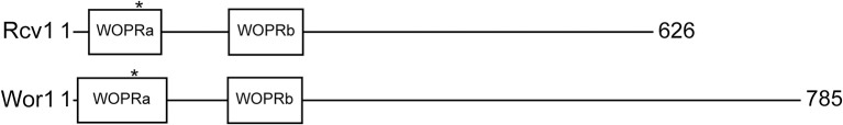
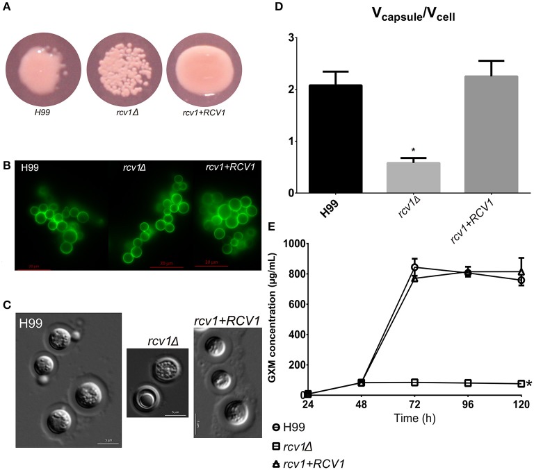
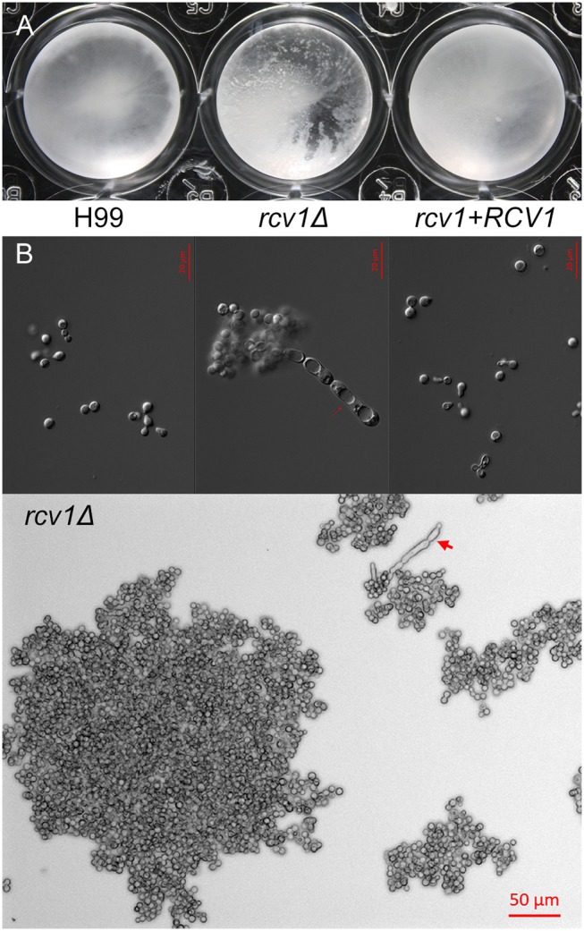
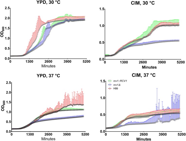
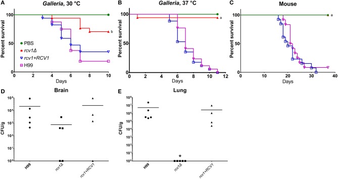
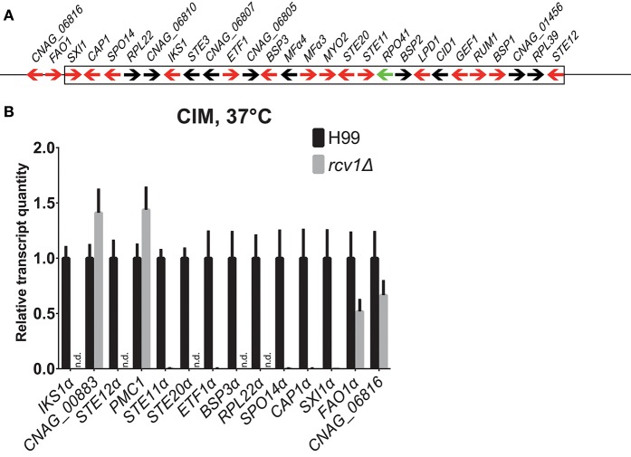
Gti1/Pac2 transcription factors occur exclusively in fungi and their roles vary according to species, including regulating morphological transition and virulence, mating and secondary metabolism. Many of these functions are important for fungal pathogenesis. We therefore hypothesized that one of the two proteins of this family in Cryptococcus neoformans, a major pathogen of humans, would also control virulence-associated cellular processes. Elimination of this protein in C. neoformans results in reduced polysaccharide capsule expression and defective cytokinesis and growth at 37°C. The mutant loses virulence in a mouse model of cryptococcal infection and retains only partial virulence in the Galleria mellonella alternative model at 30°C. We performed RNA-Seq experiments on the mutant and found abolished transcription of genes that, in combination, are known to account for all the observed phenotypes. The protein has been named Required for cytokinesis and virulence 1 (Rcv1).

Keywords: Cryptococcus neoformans; capsule; cytokinesis; transcription factor; virulence.


Figures

Figure 1
Figure 1
Schematic representation of Rcv1 (top) and Wor1 (bottom) The proteins and their features are drawn to scale. Wor1 is drawn from data of Lohse et al. (2010). The asterisks mark the position of the conserved threonine that is believed to be phosphorylated.
Figure 2
Figure 2
The rcv1Δ strain produces capsule, but fails at induction. (A) The mutant colonies appear dry on capsule-inducing solid medium at 37°C. DMEM-MOPS, 72 h. (B) GXM detection by the 2D10 mAb. The clumping is an artifact caused by the antibody, an IgM. Fluorescence. Sabouraud/MOPS, 24 h, 37°C. (C) Percoll®; observation of the capsule. The mutant has a thin, but visible capsule. DMEM-MOPS, 120 h. (D) Comparison of the mean ratios of capsule and cell volumes for cells of each strain (50 per group). DMEM/MOPS, 5 days, 37°C. *p < 0.0001, Tukey's post-test, one-way ANOVA. Bars, CI95%. (E) GXM secretion pattern, as determined by ELISA, of the mutant strain on minimal medium relative to H99 and the reconstituted strains. This plot is representative of two experiments, each one done in duplicate. Means with SDs. *p < 0.05, Dunnett's multiple comparisons test, two-way ANOVA.
Figure 3
Figure 3
The rcv1Δ strain shows heavy clumping under certain conditions. (A) Clumping of cells becomes macroscopically apparent for the mutant strain after 9 days at 37°C on Sab/MOPS. (B) Upper panel: DIC micrographs of yeast cells from the three strains after 7 days on CO2-independent medium at 37°C. Notice the elongated cells with prominent vacuoles on the mutant. Lower panel: large clump of rcv1Δ cells on same conditions. Arrows, pseudohypha-like structures.
Figure 4
Figure 4
At 37°C or on cell culture medium, the rcv1Δ mutant shows retarded growth and reaches stationary phase at lower densities. C. neoformans yeast cells were inoculated in triplicate at a density of 104 cells/mL and incubated at the indicated media and temperatures under orbital shaking and cultures had their OD600 measured every 30 min. The high dispersion in measurements observed at later time points for the mutant strain on CIM at 37°C was caused by heavy clumping of cells. The experiment is representative of two and each sample was processed in triplicate. Error bars are 95% confidence intervals (CI95%) and the same color of the curves (red, H99; blue, rcv1Δ; green, rcv1 + RCV1).
Figure 5
Figure 5
The rcv1Δ strain is hypo- or avirulent in vivo. (A–C) Survival curves in caterpillar and murine models. G. mellonella were infected with 104 yeast cells per individual, whereas BALB/C mice were infected with 2 × 10⁵ cells each. The “a” indicates statistical difference relative to H99 and rcv1+RCV1, and “b” indicates statistical difference relative to all the other groups (log-rank test, p < 0.001). (D,E) Fungal burdens in brain and lung from mice at day 12 of infection. Groups were compared using the Kruskal-Wallis test. The asterisk indicates significance (p < 0.05) relative to the other two groups. Bars, means.
Figure 6
Figure 6
The rcv1Δ strain does not cause damage to the lungs. Mice were infected as in Figure 5 (4–5 individuals per group) and lungs were harvested at day seven. (A) Normal lung. (B) H99 infection. (C) rcv1Δ infection. (D) rcv1+RCV1 infection. HE staining, bright field. Green arrowheads, five randomly selected yeast cells per field in (A,C), for illustration.
Figure 7
Figure 7
The Rcv1-GFP chimera localizes to the nucleus. The RCV1-GFP strain was grown for 16 h on YPD at 30°C before cells from the culture were applied to a microscope slide. Rcv1-GFP cells were stained with Hoechst®; 33342, showing that the nuclear stain co-localizes with the GFP signal. Bar, 5 μm. DIC, differential interference contrast microscopy. See Methods for details.
Figure 8
Figure 8
The genes the expression whereof was most strongly affected by Rcv1 deletion are clustered in the MATα locus. (A) Map of the MATα locus of H99 (not to scale) based on the latest assembly by the Broad Institute. Arrows indicate annotated ORFs. Those in red are induced in the macrophage according to the work of Heitman and colleagues (Fan et al., 2005) and repressed in the rcv1Δ strain in our RNA-Seq data. The only ORF for which data of the two studies are divergent, RPO41α, is indicated as a green arrow. The box indicates the borders of the locus and two upstream ORFs that are also repressed in the rcv1Δ are also shown. See text for details. (B) Quantitative PCR validation of RNA-Seq data for a selection of RNA-Seq differential ORFs. The plots indicate relative transcript quantitation using ACT1 as the housekeeping transcript and H99, 4 h CIM at 37°C as reference. Bars are SDs. Samples were the same used for the RNA-Seq itself plus two independent RNA extractions for each condition. N.d., not detectable (transcripts were less than 0.01% of wild-type). The y axis is linear. See text for details.

Similar articles

Cited by

References

    1. Beyhan S., Gutierrez M., Voorhies M., Sil A. (2013). A temperature-responsive network links cell shape and virulence traits in a primary fungal pathogen. PLoS Biol. 11:e1001614. 10.1371/journal.pbio.1001614 - DOI - PMC - PubMed
    1. Byrnes E. J., Bartlett K. H., Perfect J. R., Heitman J. (2011). Cryptococcus gattii: an emerging fungal pathogen infecting humans and animals. Microbes Infect. 13, 895–907. 10.1016/j.micinf.2011.05.009 - DOI - PMC - PubMed
    1. Cain C. W., Lohse M. B., Homann O. R., Sil A., Johnson A. D. (2012). A conserved transcriptional regulator governs fungal morphology in widely diverged species. Genetics 190, 511–521. 10.1534/genetics.111.134080 - DOI - PMC - PubMed
    1. Caspari T. (1997). Onset of gluconate-H+ symport in Schizosaccharomyces pombe is regulated by the kinases Wis1 and Pka1, and requires the gti1+ gene product. J. Cell Sci. 110(Pt 20), 2599–2608. - PubMed
    1. Chang Y. C., Wickes B. L., Miller G. F., Penoyer L. A., Kwon-Chung K. J. (2000). Cryptococcus neoformans STE12α Regulates Virulence but Is Not Essential for Mating. J. Exp. Med. 191, 871–882. 10.1084/jem.191.5.871 - DOI - PMC - PubMed

Publication types

LinkOut - more resources