Histological Disorganization of Spleen Compartments and Severe Visceral Leishmaniasis
- PMID: 30483481
- PMCID: PMC6243053
- DOI: 10.3389/fcimb.2018.00394
Histological Disorganization of Spleen Compartments and Severe Visceral Leishmaniasis
Abstract
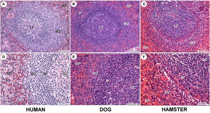
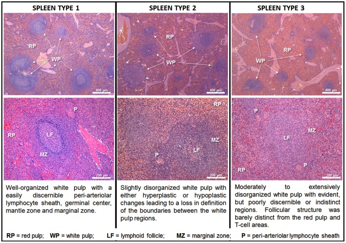
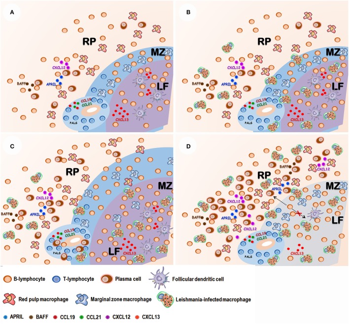
The spleen is a secondary lymphoid organ responsible for immune surveillance against blood-circulating pathogens. Absence of the spleen is associated with increased susceptibility to systemic spread and fatal infection by different pathogens. Severe forms of visceral leishmaniasis are associated with disorganization of spleen compartments where cell interactions essential for splenic immunological function take place. White pulp atrophies, secondary lymphoid follicles and marginal zones vanish, and the boundaries separating white and red pulp blur. Leukocyte populations are reduced or disappear or are replaced by plasma cells. In this paper, we review the published data on spleen disorganization in severe forms of visceral leishmaniasis and propose a histological classification to help the exchange of information among research groups.
Keywords: Leishmania infantum; spleen disorganization; spleen pathology; visceral leishmaniasis; white pulp disruption.
Figures


Similar articles
-
Low CXCL13 expression, splenic lymphoid tissue atrophy and germinal center disruption in severe canine visceral leishmaniasis.PLoS One. 2012;7(1):e29103. doi: 10.1371/journal.pone.0029103. Epub 2012 Jan 5. PLoS One. 2012. PMID: 22242159 Free PMC article.
-
Phenotypical Characterization of Spleen Remodeling in Murine Experimental Visceral Leishmaniasis.Front Immunol. 2020 Apr 15;11:653. doi: 10.3389/fimmu.2020.00653. eCollection 2020. Front Immunol. 2020. PMID: 32351510 Free PMC article.
-
Pro-Cellular Exhaustion Markers are Associated with Splenic Microarchitecture Disorganization and Parasite Load in Dogs with Visceral Leishmaniasis.Sci Rep. 2019 Sep 10;9(1):12962. doi: 10.1038/s41598-019-49344-1. Sci Rep. 2019. PMID: 31506501 Free PMC article.
-
Systemic and compartmentalized immune response in canine visceral leishmaniasis.Vet Immunol Immunopathol. 2009 Mar 15;128(1-3):87-95. doi: 10.1016/j.vetimm.2008.10.307. Epub 2008 Oct 17. Vet Immunol Immunopathol. 2009. PMID: 19054576 Review.
-
Balancing immunity and pathology in visceral leishmaniasis.Immunol Cell Biol. 2007 Feb-Mar;85(2):138-47. doi: 10.1038/sj.icb7100011. Epub 2006 Dec 5. Immunol Cell Biol. 2007. PMID: 17146466 Review.
Cited by
-
High levels of anti-Leishmania IgG3 and low CD4+ T cells count were associated with relapses in visceral leishmaniasis.BMC Infect Dis. 2021 Apr 20;21(1):369. doi: 10.1186/s12879-021-06051-5. BMC Infect Dis. 2021. PMID: 33874901 Free PMC article.
-
Bidimensional and Contrast-Enhanced Ultrasonography of the Spleen in Dogs Affected by Leishmaniosis.Animals (Basel). 2021 May 17;11(5):1437. doi: 10.3390/ani11051437. Animals (Basel). 2021. PMID: 34067930 Free PMC article.
-
Anti-Leishmania infantum Antibody-Producing Plasma Cells in the Spleen in Canine Visceral Leishmaniasis.Pathogens. 2021 Dec 17;10(12):1635. doi: 10.3390/pathogens10121635. Pathogens. 2021. PMID: 34959592 Free PMC article.
-
Lymphocyte HVEM/BTLA co-expression after critical illness demonstrates severity indiscriminate upregulation, impacting critical illness-induced immunosuppression.Front Med (Lausanne). 2023 May 25;10:1176602. doi: 10.3389/fmed.2023.1176602. eCollection 2023. Front Med (Lausanne). 2023. PMID: 37305124 Free PMC article.
-
Histone Deacetylase Inhibitors Show a Potential Leishmanicidal Effect against Leishmania braziliensis in a Mouse Infection Model and Lead to Less Toxicity than Glucantime.ACS Omega. 2025 May 6;10(19):19466-19479. doi: 10.1021/acsomega.4c11381. eCollection 2025 May 20. ACS Omega. 2025. PMID: 40415797 Free PMC article.
References
-
- Abreu C. B., de Navarro, I. T., Balarin M. R. S., Bracarense A. P. F. R. L., Marana E. R. M., Trapp S. M., et al. (2001). Aspectos clínicos, patológicos e sorológicos da toxoplasmose experimental em cães jovens. Sem. Ci. Agr. 22, 123–129. 10.5433/1679-0359.2001v22n2p123 - DOI
-
- Al-Shaebi E. M., Taib N. T., Mubaraki M. A., Hafiz T. A., Lokman M. S., Al-Ghamdy A. O., et al. . (2017). Indigofera oblongifolia leaf extract regulates spleen macrophage response during Plasmodium chabaudi infection. Saudi J. Biol. Sci. 24, 1663–1666. 10.1016/j.sjbs.2017.06.006 - DOI - PMC - PubMed
Publication types
MeSH terms
LinkOut - more resources
Full Text Sources