Review
. 2018 Nov 13:6:147.
doi: 10.3389/fcell.2018.00147. eCollection 2018.

Selective Autophagy and Xenophagy in Infection and Disease

Affiliations
Review

Selective Autophagy and Xenophagy in Infection and Disease

Vartika Sharma et al. Front Cell Dev Biol. .

Abstract

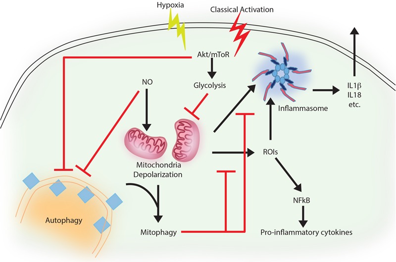
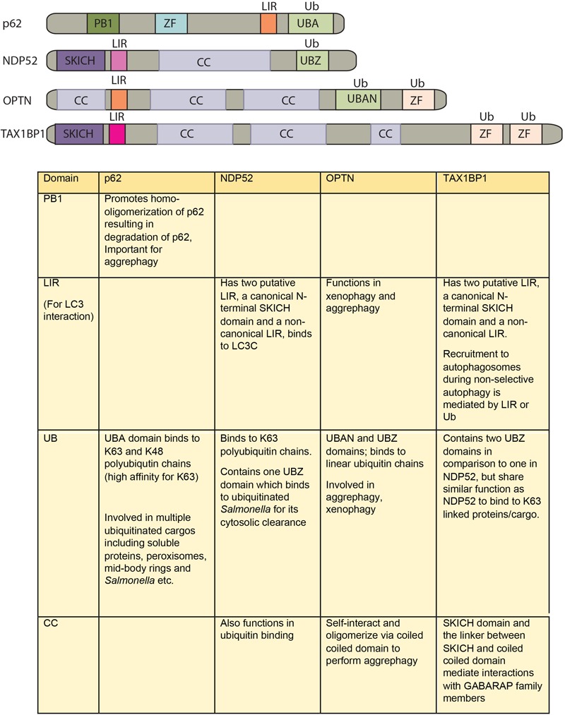
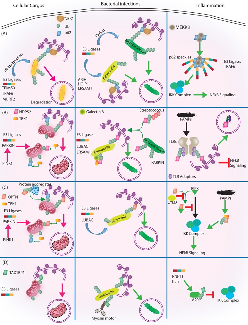
Autophagy, a cellular homeostatic process, which ensures cellular survival under various stress conditions, has catapulted to the forefront of innate defense mechanisms during intracellular infections. The ability of autophagy to tag and target intracellular pathogens toward lysosomal degradation is central to this key defense function. However, studies involving the role and regulation of autophagy during intracellular infections largely tend to ignore the housekeeping function of autophagy. A growing number of evidences now suggest that the housekeeping function of autophagy, rather than the direct pathogen degradation function, may play a decisive role to determine the outcome of infection and immunological balance. We discuss herein the studies that establish the homeostatic and anti-inflammatory function of autophagy, as well as role of bacterial effectors in modulating and coopting these functions. Given that the core autophagy machinery remains largely the same across diverse cargos, how selectivity plays out during intracellular infection remains intriguing. We explore here, the contrasting role of autophagy adaptors being both selective as well as pleotropic in functions and discuss whether E3 ligases could bring in the specificity to cargo selectivity.

Keywords: DUBs; NDP52; OPTN; TAX1BP1; inflammation; p62; ubiquitination; xenophagy.


Figures

FIGURE 1
FIGURE 1
Autophagy as an anti-inflammatory process. Activation of macrophages upon IFNγ and LPS treatment results in classically activated macrophages, which are more phagocytic, microbicidal and inflammatory in nature. During classical activation, autophagy is inhibited in a NO-dependent manner. This coincides with mitochondrial depolarization and accumulation of such depolarized mitochondria due to lack of mitophagy (since the core machinery for autophagy and mitophagy are same). Depolarized mitochondria are source of heightened reactive oxygen intermediates (ROIs) in the activated macrophages and also activates NLRP3 mediated inflammasome pathway and secretion of cytokines like IL1β and IL18. Inhibition of autophagy occurs in mToR/Akt dependent manner and inhibition of Akt signaling can alleviate the autophagy levels and control mitochondrial depolarization by directing damaged mitochondria toward mitophagy. Together, inhibition of autophagy in these cells helps achieve the pro-inflammatory phenotype, establishing the anti-inflammatory function of autophagy. Similar events also occur when macrophages are exposed to hypoxia, with the exception of NO production, which does not occur under hypoxia.
FIGURE 2
FIGURE 2
Domain structure of autophagy adaptors and their function. The following abbreviations are used for each domain: PB1, Phox and Bem1 domain; CC, coiled-coil domain; LIR, LC3-interacting region; UBA, ubiquitin-associated domain; SKICH, SKIP carboxyl homology domain; ZF/UBZ, Ubiquitin binding Zinc-finger domain; UBAN, ubiquitin binding in ABIN and NEMO domain.
FIGURE 3
FIGURE 3
Autophagy adaptors in selective autophagy, xenophagy and inflammation. Roles of different autophagy adaptors are highlighted here in the context of selective autophagy of intracellular cargos, xenophagy and inflammation. (A) p62/SQSTM1: E3 ligases TRIM 50, TRAF 6, MURF 2 ubiquitinate the intracellular cargos, followed by their recognition by autophagy adapter p62 and NBR1. Adaptors then target the cargo to autophagosome for subsequent degradation. During bacterial infection, p62 adaptor protein recognizes targets that are tagged by E3 ligases like ARIH, HOIP1, LRSAM1 (which act on Salmonella), after which p62 and NBR1 are recruited to the bacteria and targets Salmonella for autophagic degradation. In case of M. tuberculosis so far only Parkin has been shown to act as E3 ligase leading to K63 ubiquitination, p62-NBR1 recruitment and targeting of M. tuberculosis to autophagosomes. Finally, p62 is directly implicated in regulating inflammation. The PB1 domains of p62 homo and heterodimerize while interacting with MEKKK3. This complex further co-localizes to TRAF 6 oligomers, forming what is known as p62 speckles. This complex then phosphorylates and ubiquitinates IKK complex, inhibiting NFκB signaling. (B) NDP52/CALCOCO2: Upon mitochondrial damage the recruitment of PINK1/PARKIN E3 ligase help in ubiquitinating mitochondria and activating TBK1, which subsequently phosphorylate both NDP52 and p62 Ubiquitination of mitochondria and tagging with p62 and NDP52 helps in targeting mitochondria to autophagosome. During Salmonella infection, LUBAC complex and LSRAM1 E3 ligases ubiquitinate the bacteria. Phosphorylation of NDP52 by TBK1, help tagging of the bacteria with NDP52. Here cytosolic Galectin 8 also takes part in the process and interacts with NDP52. For recruitment of M. tuberculosis parkin mediates ubiquitination of M. tuberculosis. NDP52 is also recruited to M. tuberculosis and targets it to autophagosome. Rab35 and NDP52 also mediate targeting of Streptoccocus to autophagosomes. Bacterial PAMPS are recognized by TLR followed by recruitment of TLR adapters. The TLR adaptors get ubiquitinated and recognized by autophagy adapter NDP52. Along with the TLR adaptors, autophagy adaptors undergo degradation via autophagosome maturation called adaptophagy and controls inflammation. (C) OPTN: OPTN acts in mitophagy in a manner very similar to NDP52 where PINK1 and PARKIN E3 ligases are activated and recruited to mitochondria for ubiquitination. Ubiquitinated mitochondria are recognized by OPTN for targeting them to autophagosomes. In addition to mitophagy for degrading intracellular cargo OPTN performs aggrephagy as well. During bacterial infections, OPTN’s role has been shown in the context of LUBAC complex mediated K63 and M1 polyubiquitination of Salmonella. Upon recruitment, OPTN targets the bacterium to autophagosomes for degradation. OPTN is also shown to inhibit IKK complex by interacting with RIPK. Interaction of OPTN with a deubiquitinating enzyme called CYLD, which deubiquitinates OPTN and RIPK allows RIPK mediated activation of IKK complex and inflammation. Similarly, PAMPs can activate TBK1, which gets autophosphorylated and subsequently binds to TBK binding domain of OPTN to alleviate inflammation. (D) TAX1BP1/CALCOCO3: Similar, to other autophagy adapters like NDP52 and OPTN TAX1BP1 also performs mitophagy where E3 ligases are not very well known, however, Parkin is the most likely candidate. In Salmonella, TAX1BP1 interacts with myosin motor VI and induces autophagosome and lysosome fusion subsequently helping in xenophagy of polyubiquitinated Salmonella. TAX1BP1 interacts with A20, an NFκB inhibitor to control inflammation via inhibiting IKK complex. Here, RNF 11 and Itch E3 ligases helps recruitment of TAX1BP1 to A20 for autophagic targeting. While we have used very selected examples to highlight the functional overlaps between different autophagy adaptors during selective autophagy, it must be noted that inhibition of selective autophagy like mitophagy also contributes to inflammation. This figure therefore showcases the complex regulatory events and points toward the existing lacunae in our understanding of selective autophagy, especially in the context of bacterial infection and inflammation.

References

    1. Arasaki K., Mikami Y., Shames S. R., Inoue H., Wakana Y., Tagaya M. (2017). Legionella effector Lpg1137 shuts down ER-mitochondria communication through cleavage of syntaxin 17. Nat. Commun. 8:15406. 10.1038/ncomms15406 - DOI - PMC - PubMed
    1. Ashida H., Kim M., Schmidt-Supprian M., Ma A., Ogawa M., Sasakawa C. (2010). A bacterial E3 ubiquitin ligase IpaH9.8 targets NEMO/IKKgamma to dampen the host NF-kappaB-mediated inflammatory response. Nat. Cell Biol. 12 66–73; sup pp 1–9. 10.1038/ncb2006 - DOI - PMC - PubMed
    1. Bach H., Papavinasasundaram K. G., Wong D., Hmama Z., Av-Gay Y. (2008). Mycobacterium tuberculosis virulence is mediated by PtpA dephosphorylation of human vacuolar protein sorting 33B. Cell Host Microbe 3 316–322. 10.1016/j.chom.2008.03.008 - DOI - PubMed
    1. Bah A., Lacarriere C., Vergne I. (2016). Autophagy-Related proteins target ubiquitin-free mycobacterial compartment to promote killing in macrophages. Front. Cell Infect Microbiol. 6:53. 10.3389/fcimb.2016.00053 - DOI - PMC - PubMed
    1. Barnett T. C., Liebl D., Seymour L. M., Gillen C. M., Lim J. Y., Larock C. N., et al. (2013). The globally disseminated M1T1 clone of group A Streptococcus evades autophagy for intracellular replication. Cell Host Microbe 14 675–682. 10.1016/j.chom.2013.11.003 - DOI - PMC - PubMed