

doi: 10.3389/fcell.2018.00150.
eCollection 2018.
Role of Metabolism in Hepatic Stellate Cell Activation and Fibrogenesis
Affiliations
- PMID: 30483502
- PMCID: PMC6240744
- DOI: 10.3389/fcell.2018.00150
Item in Clipboard
Role of Metabolism in Hepatic Stellate Cell Activation and Fibrogenesis
Front Cell Dev Biol.
.
Abstract
Activation of hepatic stellate cell (HSC) involves the transition from a quiescent to a proliferative, migratory, and fibrogenic phenotype (i.e., myofibroblast), which is characteristic of liver fibrogenesis. Multiple cellular and molecular signals which contribute to HSC activation have been identified. This review specially focuses on the metabolic changes which impact on HSC activation and fibrogenesis.
Keywords: fibroblast; glutaminolysis; glycolysis; liver fibrosis; metabolic.
Figures
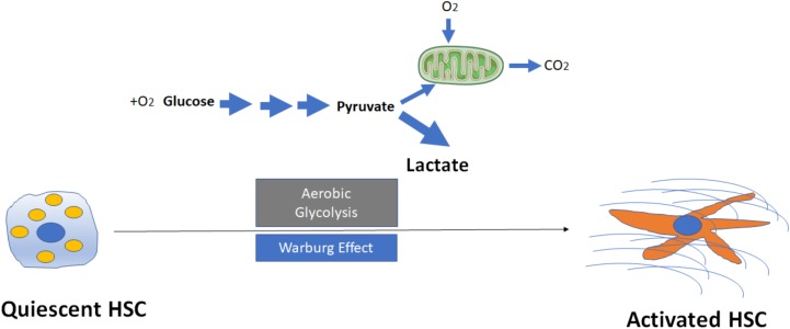
Activation of hepatic stellate cells (HSCs) through induction of aerobic glycolysis (Warburg effect). The transformation of glucose to lactate during HSC activation even when amounts of oxygen are available, leads to accumulation of intracellular lactate. Mitochondria may remain functional and some oxidative phosphorylation continue in cells. Aerobic glycolysis is less efficient than oxidative phosphorylation for generating adenosine 5′-triphosphate (ATP), which suggests that metabolites (for example, lactate) generated by aerobic glycolysis may have a more important role in the regulation of cellular functions than simply energy production during HSC activation.
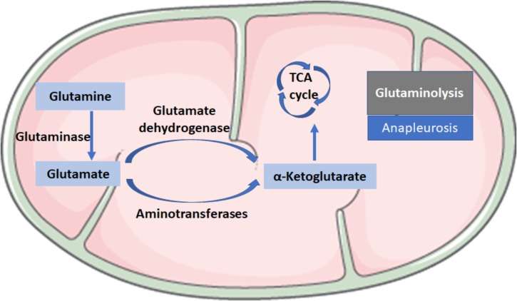
Biochemical reactions in glutaminolysis. Glutaminolysis is the conversion of glutamine (Gln) to α-ketoglutarate (α-KG) and consists of two reactions: the first reaction is catalyzed by the glutaminase (GLS), which converts Gln into glutamate (Glu) by losing an amino group; the second step consists of the conversion of Glu to α-KG, a critical intermediate in the tricarboxylic acid (TCA) cycle, which is catalyzed by glutamate dehydrogenase or aminotransferases.
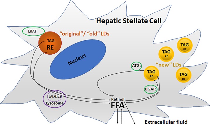
Two different metabolic pools of lipid droplets (LDs) in activated HSCs. The “original”/“old” LDs (depicted in brown), are located predominately round the nucleus, and contains predominantly triacylglycerol (TAG) and retinyl ester (RE), as well as retinol acyltransferase (LRAT). Lysosomal acid lipase (LAL/Lipa) is involved in the degradation of the “original”/“old” LDs in the lysosome during activation. The “new” LDs (depicted in yellow) which are smaller than “old” LDs, contain less REs but are enriched in TAGs, and are located in the periphery of the cells. Diacylglycerol O-acyltransferase 1 (DGAT1) and adipose triglyceride lipase (ATGL) are involved in the synthesis and breakdown of these newly synthesized TAGs, respectively.
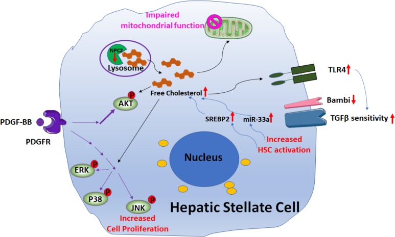
Signaling pathways involved in free cholesterol (FC) accumulation mediated HSC activation. Downregulation of Niemann–Pick type C2 protein (NPC2) results in FC accumulation and enhances platelet-derived growth factor BB (PDGF-BB)-induced HSC proliferation by extracellular signal-regulated kinases (ERKs), p38, c-Jun N-terminal kinases (JNK), and protein kinase B (AKT) phosphorylation. In addition, the mitochondrial respiration function is impaired. FC accumulation also increases Toll-like receptor 4 protein (TLR4) expression, thereby sensitizing cells to TGF-β-induced activation through down-regulation of TGFβ-pseudoreceptor Bambi. Along with HSC activation, subsequent upregulation of both sterol regulatory element-binding protein 2 (SREBP2) and miR-33a signaling leads to further FC accumulation and exaggerates liver fibrosis in a positive feedforward loop.
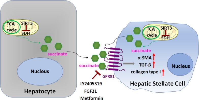
Role of succinate in HSC activation. Succinate, an intermediate in the TCA cycle, functions as a paracrine signal between hepatocytes and HSCs, through binding and activation of its cognate G protein-coupled receptor 91 (GPR91), which resulted in upregulation of fibrogenic markers alpha-smooth muscle actin (α-SMA), transforming growth factor β (TGF-β), and collagen type I. Sirtuin 3 (SIRT3), a NAD+-dependent protein deacetylase, predominantly localized in the mitochondrial matrix, is a key regulator of dehydrogenase (SDH) activity. The SIRT3-SDH-GPR91 axis regulates HSC activation. Repression of succinate-GPR91 signaling by LY2405319, an analog of fibroblast growth factor 21 (FGF21), as well as metformin inhibits HSC activation.
Similar articles
-
Cooperation of liver cells in health and disease.Adv Anat Embryol Cell Biol. 2001;161:III-XIII, 1-151. doi: 10.1007/978-3-642-56553-3. Adv Anat Embryol Cell Biol. 2001. PMID: 11729749 Review.
-
Molecular interplays in hepatic stellate cells: apoptosis, senescence, and phenotype reversion as cellular connections that modulate liver fibrosis.Cell Biol Int. 2017 Sep;41(9):946-959. doi: 10.1002/cbin.10790. Epub 2017 Jul 20. Cell Biol Int. 2017. PMID: 28498509 Review.
-
Overexpression of c-myc in hepatocytes promotes activation of hepatic stellate cells and facilitates the onset of liver fibrosis.Biochim Biophys Acta. 2013 Oct;1832(10):1765-75. doi: 10.1016/j.bbadis.2013.06.001. Epub 2013 Jun 12. Biochim Biophys Acta. 2013. PMID: 23770341
-
Therapeutic effects of serum extracellular vesicles in liver fibrosis.J Extracell Vesicles. 2018 Apr 17;7(1):1461505. doi: 10.1080/20013078.2018.1461505. eCollection 2018. J Extracell Vesicles. 2018. PMID: 29696080 Free PMC article.
-
SIRT1 antagonizes liver fibrosis by blocking hepatic stellate cell activation in mice.FASEB J. 2018 Jan;32(1):500-511. doi: 10.1096/fj.201700612R. Epub 2017 Sep 26. FASEB J. 2018. PMID: 28970250
Cited by
-
A novel minimally invasive OFM technique with orthotopic transplantation of hUC-MSCs and in vivo monitoring of liver metabolic microenvironment in liver fibrosis treatment.Stem Cell Res Ther. 2021 Oct 9;12(1):534. doi: 10.1186/s13287-021-02599-w. Stem Cell Res Ther. 2021. PMID: 34627378 Free PMC article.
-
Fibrogenic Pathways in Metabolic Dysfunction Associated Fatty Liver Disease (MAFLD).Int J Mol Sci. 2022 Jun 23;23(13):6996. doi: 10.3390/ijms23136996. Int J Mol Sci. 2022. PMID: 35805998 Free PMC article. Review.
-
Mitofusin-2 Restrains Hepatic Stellate Cells' Proliferation via PI3K/Akt Signaling Pathway and Inhibits Liver Fibrosis in Rats.J Healthc Eng. 2022 Jan 17;2022:6731335. doi: 10.1155/2022/6731335. eCollection 2022. J Healthc Eng. 2022. PMID: 35083025 Free PMC article.
-
Gut microbiota mediated molecular events and therapy in liver diseases.World J Gastroenterol. 2020 Dec 28;26(48):7603-7618. doi: 10.3748/wjg.v26.i48.7603. World J Gastroenterol. 2020. PMID: 33505139 Free PMC article. Review.
-
Treatment of liver fibrosis: Past, current, and future.World J Hepatol. 2023 Jun 27;15(6):755-774. doi: 10.4254/wjh.v15.i6.755. World J Hepatol. 2023. PMID: 37397931 Free PMC article. Review.
References
-
- Ajat M., Molenaar M., Brouwers J. F. H. M., Vaandrager A. B., Houweling M., Helms J. B. (2017). Hepatic stellate cells retain the capacity to synthesize retinyl esters and to store neutral lipids in small lipid droplets in the absence of LRAT. Biochim. Biophys. Acta 1862 176–187. 10.1016/j.bbalip.2016.10.013 - DOI - PubMed
LinkOut - more resources
Full Text Sources