Review
. 2018 Nov 13:5:91.
doi: 10.3389/fmolb.2018.00091. eCollection 2018.

The PII-NAGK-PipX-NtcA Regulatory Axis of Cyanobacteria: A Tale of Changing Partners, Allosteric Effectors and Non-covalent Interactions

Affiliations
Review

The PII-NAGK-PipX-NtcA Regulatory Axis of Cyanobacteria: A Tale of Changing Partners, Allosteric Effectors and Non-covalent Interactions

Alicia Forcada-Nadal et al. Front Mol Biosci. .

Abstract

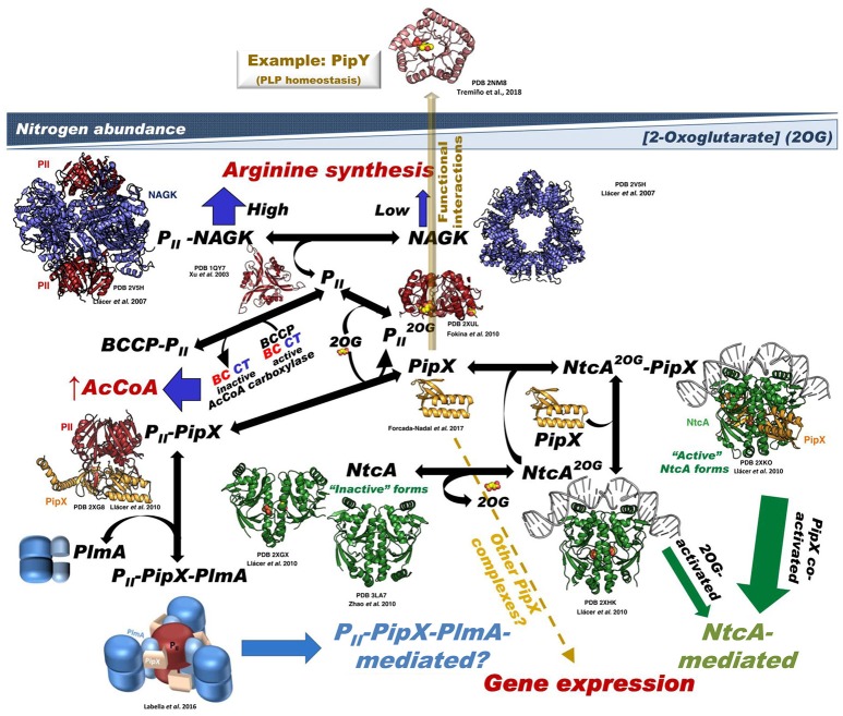
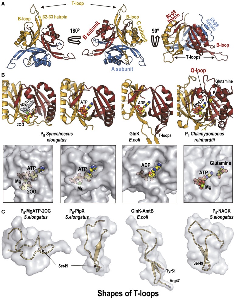
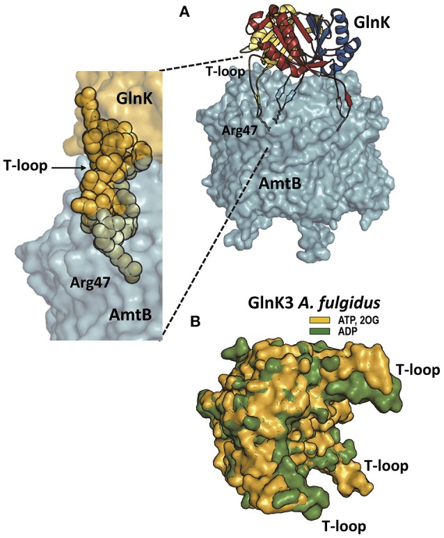
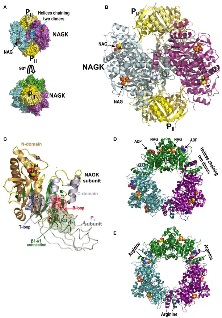
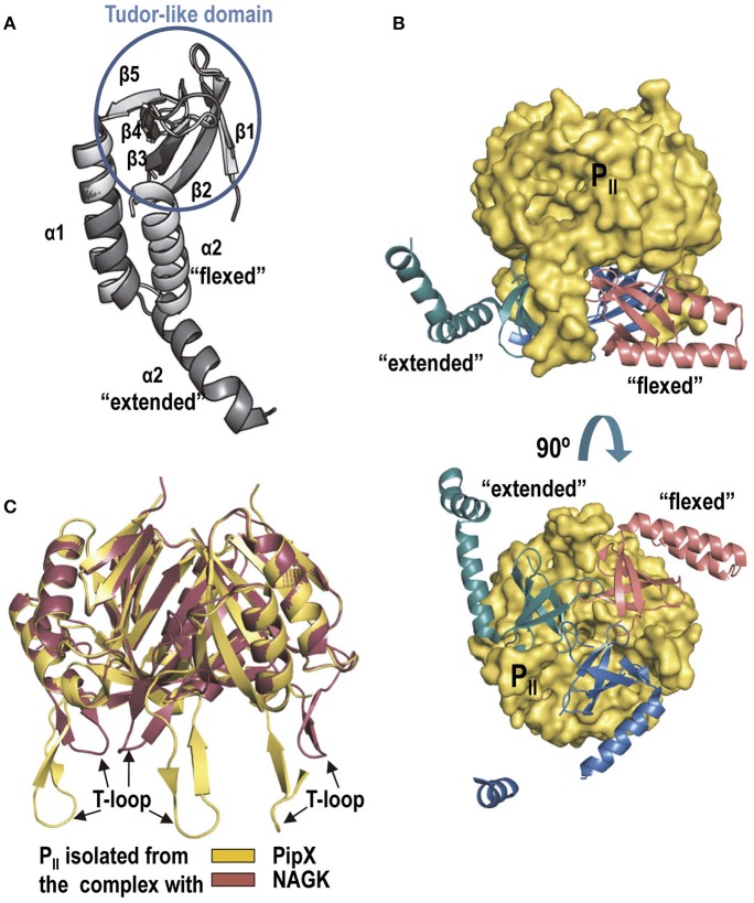
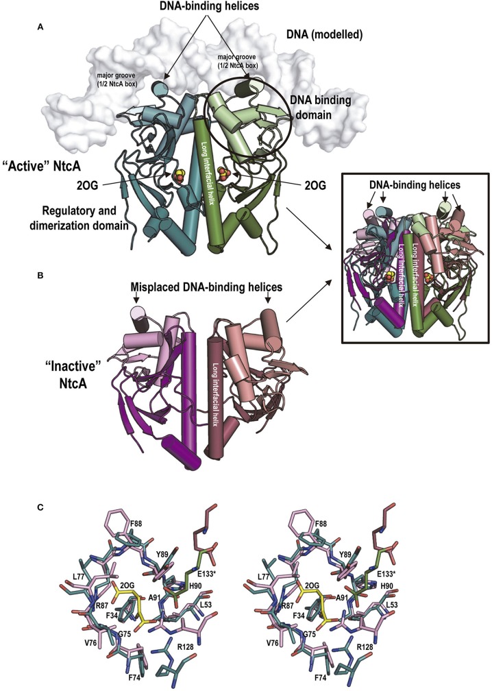
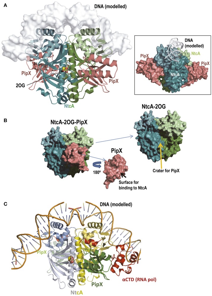
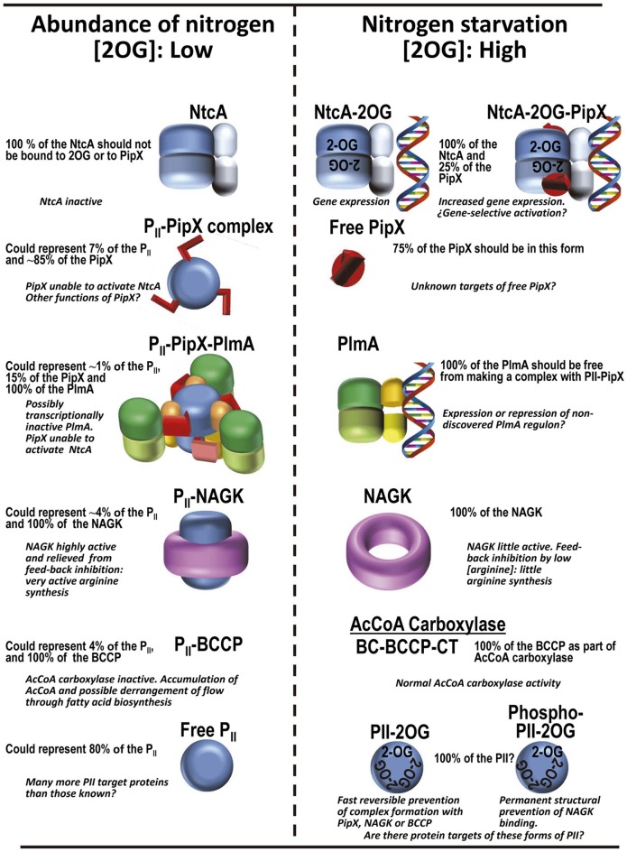
PII, a homotrimeric very ancient and highly widespread (bacteria, archaea, plants) key sensor-transducer protein, conveys signals of abundance or poorness of carbon, energy and usable nitrogen, converting these signals into changes in the activities of channels, enzymes, or of gene expression. PII sensing is mediated by the PII allosteric effectors ATP, ADP (and, in some organisms, AMP), 2-oxoglutarate (2OG; it reflects carbon abundance and nitrogen scarcity) and, in many plants, L-glutamine. Cyanobacteria have been crucial for clarification of the structural bases of PII function and regulation. They are the subject of this review because the information gathered on them provides an overall structure-based view of a PII regulatory network. Studies on these organisms yielded a first structure of a PII complex with an enzyme, (N-acetyl-Lglutamate kinase, NAGK), deciphering how PII can cause enzyme activation, and how it promotes nitrogen stockpiling as arginine in cyanobacteria and plants. They have also revealed the first clear-cut mechanism by which PII can control gene expression. A small adaptor protein, PipX, is sequestered by PII when nitrogen is abundant and is released when is scarce, swapping partner by binding to the 2OG-activated transcriptional regulator NtcA, co-activating it. The structures of PII-NAGK, PII-PipX, PipX alone, of NtcA in inactive and 2OG-activated forms and as NtcA-2OG-PipX complex, explain structurally PII regulatory functions and reveal the changing shapes and interactions of the T-loops of PII depending on the partner and on the allosteric effectors bound to PII. Cyanobacterial studies have also revealed that in the PII-PipX complex PipX binds an additional transcriptional factor, PlmA, thus possibly expanding PipX roles beyond NtcA-dependency. Further exploration of these roles has revealed a functional interaction of PipX with PipY, a pyridoxal-phosphate (PLP) protein involved in PLP homeostasis whose mutations in the human ortholog cause epilepsy. Knowledge of cellular levels of the different components of this PII-PipX regulatory network and of KD values for some of the complexes provides the basic background for gross modeling of the system at high and low nitrogen abundance. The cyanobacterial network can guide searches for analogous components in other organisms, particularly of PipX functional analogs.

Keywords: NtcA structure and complexes; PII complexes; PipX complexes; PlmA; gene expression regulation; nitrogen regulation; protein structure; signaling.


Figures

Figure 1
Figure 1
Summary of the PII-PipX-NtcA network of S. elongatus. The network illustrates its different elements and complexes depending on nitrogen abundance (inversely related to 2OG level) and the structures of the macromolecules and complexes formed (when known). For PlmA (dimer in darker and lighter blue hues for its dimerization and DNA-binding domains, respectively) and its complex the architectural coarse model proposed (Labella et al., 2016) is shown, with the C-terminal helices of PipX (schematized in the extended conformation) pink-colored and the two PII molecules in dark red. The DNA complexed with NtcA and with NtcA-PipX is modeled from the structure of DNA-CRP (Llácer et al., 2010), since no DNA-NtcA structure has been reported. BCCP, biotin carboxyl carrier protein of bacterial acetyl CoA carboxylase (abbreviated AcCoA carboxylase); the other two components of this enzyme, biotin carboxylase and carboxyl transferase are abbreviated BC and CT, respectively. No structural model of BCC has been shown because the structure of this component has not been determined in S. elongatus and also because the structures of this protein from other bacteria lack a disordered 77-residue N-terminal portion that could be highly relevant for interaction with PII. The yellow broken arrow highlights the possibility of further PipX interactions not mediated by NtcA or PII-PlmA resulting in changes in gene expression (Espinosa et al., 2014). The solid semi-transparent yellowish arrow emerging perpendicularly from the flat network symbolizes the possibility of functional interactions of PipX not mediated by physical contacts between the macromolecules involved in the interaction, giving as an example the functional interaction with PipY. Its position outside the network tries to express the different type of interaction (relative to the physical contacts shown in the remainder of the network) as well as to place it outside the field of 2OG concentrations.
Figure 2
Figure 2
The PII protein. (A) Overall view in cartoon representation of S. elongatus PII along its three-fold axis from the PII flat face (left), from its convex side (middle), or with the three-fold axis vertical and the flat surface down (right). The structure corresponds to Protein Database (PDB) file 1QY7 (Xu et al., 2003). Each subunit in the trimer is colored differently. Some relevant traits are highlighted. (B) The PII allosteric sites shown in cartoon representation (top) and in semi-transparent zoomed surface representation (bottom) in approximately the same pose as in the cartoon representation. For clarity, in the cartoon representations only the two subunits forming each site are shown, in the same colors as in (A). Ligands are shown in sticks and balls representation, with atoms of C, O, N, P, and Mg in yellow, red, blue, orange, and green, respectively, except in the leftmost cartoon figure in which all the atoms of ATP are pale gray to highlight the bound 2OG (colored). Note in the corresponding panel of the bottom row that MgATP and 2OG are nearly fully buried in the PII molecule. The organisms from which the PII derive are indicated in the figure. The two panels to the left belong to isolated S. elongatus PII (PDB file 2XZW; Fokina et al., 2010a); the third panels illustrate E. coli GlnK taken from its complex with AmtB (PDB 2NUU, Conroy et al., 2007); the rightmost panels show Chlamydomonas reinhardtii PII, taken from its complex with Arabidopsis thaliana NAGK (PDB 4USJ; Chellamuthu et al., 2014). (C) Illustration of different shapes of the T-loops found in distinct complexes with allosteric effectors or with partner proteins. The T-loop is shown in cartoon representation, within a semi-transparent surface representation as if this loop were isolated from the remainder of PII and from the protein partner in the complex. In the third panel, the side chain of Arg47 of E. coli GlnK is represented in sticks, given its importance for inhibiting the AmtB channel. Taken, from left to right, from: S. elongatus PII with MgATP and 2OG bound (PDB file 2XZW; Fokina et al., 2010a); S. elongatus PII-PipX (PDB 2XG8; Llácer et al., 2010); E. coli GlnK-AmtB complex (PDB 2NUU, Conroy et al., 2007); and PII-NAGK complex (PDB 2V5H; Llácer et al., 2007).
Figure 3
Figure 3
PII proteins and the ammonia channel. (A) The structure (PDB file 2NUU; Conroy et al., 2007) of the E. coli complex of GlnK (a PII protein in charge of ammonia channel regulation) and the ammonia channel AmtB is shown to the right, whereas the zoom to the lower left shows only a part of the complex, to highlight the interaction of one T-loop with one channel. AmtB is in semi-transparent surface representation. GlnK is in the main figure in cartoon representation with each subunit colored differently, with the side-chain of the T-loop residue Arg47 shown in sticks representation. In the zoomed image GlnK is shown in surface representation in yellow with the T-loop residues highlighted in space-filling representation, illustrating the fact that the side-chain of Arg47 is the element getting deep into the channel and blocking it. (B) Super-imposition of the structures of Archeoglobus fulgidus GlnK3 (one of the three PII proteins of the GlnK type in this archaeon; Maier et al., 2011) with ADP bound (green; PDB file code 3TA1) or with ATP and 2OG bound (yellow; PDB 3TA2) to illustrate how 2OG binding fixes the T-loops in an outwards-flexed position (relative to the positions without 2OG) that would be inappropriate for fitting the topography of the entry chambers to the three ammonia channels in trimeric Amt (the ammonia channel in this organism).
Figure 4
Figure 4
PII-NAGK complex and active and arginine-inhibited NAGK. (A) The PII-NAGK complex of S. elongatus (PDB 2V5H; Llácer et al., 2010). Surface representations of the complex formed by two PII trimers (yellow) capping on both ends the doughnut-like NAGK hexamer (trimer of dimers; each dimer in a different color). The three-fold axis is vertical (top) or perpendicular to the page (bottom). Figure of J.L. Llácer and V. Rubio taken from Chin (2008). Reprinted with permission from AAAS (B). Cartoon representation of the S. elongatus PII-NAGK complex after removing the back NAGK dimer for clarity. The three-fold symmetry axis is vertical. Reprinted from Current Opinion in Structural Biology, 18, Llácer et al., Arginine and nitrogen storage, 673–681, 2008, with permission from Elsevier. (C) PII subunit-NAGK subunit contacts. PII, NAGK, and NAG are shown as strings, ribbons, and spheres, respectively. The contacting parts of the T-loop, B-loop, and β1–α1 connection, including some interacting side chains (in sticks), are blue, red, and green, respectively. The surfaces provided by these elements form meshworks of the same colors. The NAGK central β-sheet is green, and other β-strands and the α-helices are brownish and grayish for N- and C-domains, respectively. Some NAGK elements and PII residues are labeled. This figure and its legend reproduce with some modifications a figure and its legend of Llácer et al. (2007). The crystal structure of the complex of PII and acetylglutamate kinase reveals how PII controls the storage of nitrogen as arginine. Copyright (2007) National Academy of Sciences. (D,E), active and inactive conformations, respectively, of hexameric arginine-inhibitable NAGK. The active form is from a crystal of the enzyme from Pseudomonas aeruginosa (PDB 2BUF) while the inactive form is from the Thermotoga maritima enzyme (PDB 2BTY) (Ramón-Maiques et al., 2006). Note that the inactive form is widened relative to the active form, and that it has arginine sitting on both sides of each interdimeric junction. In the active form the nucleotide (in this case the product ADP rather than the substrate ATP) and NAG sit one in each domain of individual subunits. The NAGK observed in the PII-NAGK complex is in the active form, being stabilized in this form by its contacts with PII.
Figure 5
Figure 5
PipX and the PII-PipX complex. (A) The structures of PipX in the flexed conformation and in one of the two extended conformations observed in the S. elongatus PII-PipX complex are shown (extracted from PDB 2XG8; Llácer et al., 2010). Note that the major difference between the two conformations is the large movement of the C-terminal helix around its flexible linker (Forcada-Nadal et al., 2017). The same flexed conformation was observed in the complex with NtcA (see below) and agrees with the data of structural NMR studies on isolated PipX (Forcada-Nadal et al., 2017). The elements of the Tudor-like domain are encircled in a blue circumference. (B) The PII-PipX complex of S. elongatus viewed with the three-fold axis of PII vertical (top) or in a view along this axis, looking at the flat face of PII (bottom). (C) Superimposition of S. elongatus PII in the complex with PipX and in that with NAGK. The changes in the T-loops are very patent.
Figure 6
Figure 6
NtcA structure, 2OG binding to it and associated conformational changes. (A,B), structures of “active” (A) and “inactive” (B) S. elongatus NtcA (PDB files 2XHK and 2XGX, respectively) (Llácer et al., 2010). The two subunits of each dimer are in different colors, with the DNA-binding domains in a lighter hue than the regulatory domain of the same subunit. In the cartoon representation used, helices are shown as cylinders to illustrate best the changes in position of the DNA binding helices and of the long interfacial helices (labeled) upon activation. Bound 2OG is shown in “active” NtcA (in spheres representation, with C and O atoms colored yellow and red, respectively). The DNA, in surface representation in white, has been modeled from the CRP-DNA structure (for details see Llácer et al., 2010). The inset superimposes the “active” and “inactive” forms colored as in the main figure to illustrate the magnitude of the changes. (C) Stereo view of sticks representation of the 2OG site residues of the “active” (green) and “inactive” (raspberry) forms of NtcA. The 2OG bound to the “active” form is distinguished by its yellow C atoms. Note that only two residues, both 2OG-interacting and highly polar, experience large changes in their positions between the inactive and the active forms: Arg128 from the long interfacial helix of the subunit that provides the bulk of the residues of the site, and Glu133 from the interfacial helix of the other subunit. They are believed to trigger the changes in the relations between the two interfacial helices that result in NtcA “activation”.
Figure 7
Figure 7
The NtcA-PipX complex. (A) Structure of the complex of S. elongatus NtcA and PipX, with DNA modeled in semi-transparent surface representation (Llácer et al., 2010). The projection of NtcA differs somewhat from that in Figure 6A to allow visualization of both PipX molecules, illustrating the fact that the Tudor-like domain is the part of PipX that binds to NtcA. Note that the two PipX molecules (the asymmetric unit contained an entire complex with two PipX molecules, PDB file 2XKO) are in the “flexed” conformation, that the flexed helices protrude away from the NtcA molecule and that they do not contact the modeled bound DNA (main figure and inset). The inset represents the same complex in a different orientation (viewed approximately along the DNA) and with all elements in surface representation except the DNA (shown in cartoon) to better visualize the protrusion in large part due to the flexed helices of both PipX molecules. (B) Deconstruction of the NtcA-PipX complex to show in surface representation the crater at the NtcA surface where PipX binds, and the surface of Pip X used in this binding. (C) A model based on CRP (Llácer et al., 2010) for the complex of the NtcAPipX complex with DNA and with the C-terminal domain of the α-subunit of RNA polymerase (αCTD), to show that the C-terminal helices of PipX could reach this part of the polymerase. In this figure the C-terminal helix of NtcA is colored red because it has no counterpart in CRP and is involved in the interactions with PipX.
Figure 8
Figure 8
Protein complexes of the PII regulatory system in S. elongatus according to availability of ammonium in the cell, and their corresponding functional consequences. The frequencies of the different chains in the various forms are based on the levels of the proteins in the cell found in proteomic studies (Table 1). The PII trimer has been colored blue, PipX and its C-terminal helices are red, PlmA dimers have their DNA binding domains yellow or orange and their dimerization domains in two hues of green, NAGK is shown as a purple crown, and the regulatory and DNA binding domains of NtcA are given dark and light shades of blue, respectively.

Similar articles

Cited by

References

    1. Beez S., Fokina O., Herrmann C., Forchhammer K. (2009). N-acetyl-L-glutamate kinase (NAGK) from oxygenic phototrophs: PII signal transduction across domains of life reveals novel insights in NAGK control. J. Mol. Biol. 389, 748–758. 10.1016/j.jmb.2009.04.053 - DOI - PubMed
    1. Berg O. G., von Hippel P. H. (1988). Selection of DNA binding sites by regulatory proteins. II. The binding specificity of cyclic AMP receptor protein to recognition sites. J. Mol. Biol. 200, 709–723. - PubMed
    1. Burillo S., Luque I., Fuentes I. (2004). Interactions between the nitrogen signal transduction protein PII and N-acetyl glutamate kinase in organisms that perform oxygenic photosynthesis. J. Bacteriol. 186, 3346–3354. 10.1128/JB.186.11.3346-3354.2004 - DOI - PMC - PubMed
    1. Camargo S., Valladares A., Forchhammer K., Herrero A. (2014). Effects of PipX on NtcA-dependent promoters and characterization of the cox3 promoter region in the heterocyst-forming cyanobacterium Anabaena sp. PCC 7120. FEBS Lett. 588, 1787–1794. 10.1016/j.febslet.2014.03.039 - DOI - PubMed
    1. Cantos C., Labella J. I., Espinosa E., Contreras A. (2018). The nitrogen regulator PipX acts in cis to prevent operon polarity. Environ. Microbiol. Rep. 10.1111/1758-2229.12688 - DOI - PubMed