

Esophageal tuberculosis: Uncommon of common
- PMID: 30483561
- PMCID: PMC6207044
- DOI: 10.1002/jgh3.12043
Esophageal tuberculosis: Uncommon of common
Abstract
Background and aim: Esophageal involvement in tuberculosis (TB) is rare and is usually secondary. Data on esophageal TB are scarce. We aimed to analyze clinical and endoscopic features and outcomes of treatment in esophageal TB.
Methods: We retrospectively identified patients with esophageal TB from January 2014 to December 2016 at GB Pant Hospital. Well-defined granuloma with or without caseation and/or acid-fast bacilli on staining either from esophageal biopsy or the adjacent mediastinal lymph node fine-needle aspiration cytology (FNAC) specimen, along with clinical features and response to antitubercular therapy (ATT), were collectively considered to diagnose definite TB. Treatment received and response to therapy were documented and analyzed.
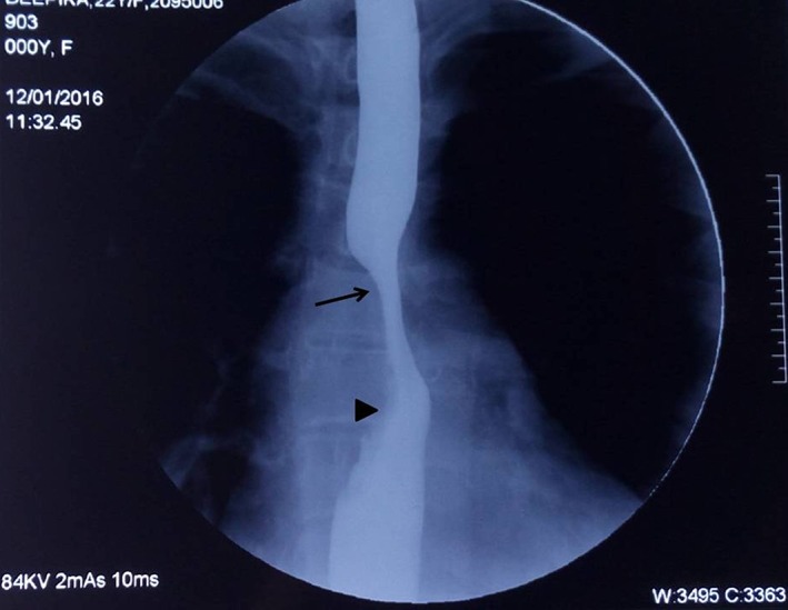
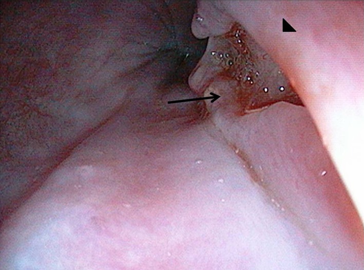
Results: A total of 19 patients had definite esophageal TB, and the median age of patients was 39 years (14-65 years) and 10 (52.6%) patients were female. The most common presenting symptom was dysphagia (n = 16, 84%) followed by odynophagia (n = 8, 42%). On endoscopy, the mid-esophagus was the most common site of involvement, and findings included ulcers (n = 17), elevated lesions (n = 9), and fistulae (n = 4) in patients. The mediastinal lymphadenopathy was present in all patients, with parenchymal lesions seen in three patients. The endoscopic mucosal biopsies were diagnostic in 11 patients, and in the remaining 8 patients, endoscopic ultrasound-guided FNAC from the mediastinal lymph nodes was diagnostic. A total of 18 patients completely responded to ATT, and 1 patient had partial response with persistent fistulae requiring additional treatment.
Conclusion: Esophagus involvement is rare in TB; endoscopic mucosal biopsy and EUS-guided FNAC is diagnostic, and the response to ATT is excellent.
Keywords: endoscopic ultrasound; endoscopy: upper gastrointestinal; esophagus; fistula; tuberculosis.
Figures


References
-
- Rubin SA. Tuberculosis. Captain of all these men of death. Radiol. Clin. North Am. 1995; 33: 619–39. - PubMed
-
- Lockard LB. Esophageal tuberculosis. A critical review. Laryngoscope. 1913; 23: 561–84.
-
- Carr DT, Spain DM. Tuberculosis in a carcinoma of the esophagus. Am. Rev. Tuberc. 1942; 46: 346–9.
LinkOut - more resources
Full Text Sources
