

Chrysomya chani Kurahashi (Diptera: Calliphoridae), a blow fly species of forensic importance: morphological characters of the third larval instar and a case report from Thailand
- PMID: 30483655
- PMCID: PMC6197134
- DOI: 10.1080/20961790.2018.1428078
Chrysomya chani Kurahashi (Diptera: Calliphoridae), a blow fly species of forensic importance: morphological characters of the third larval instar and a case report from Thailand
Abstract
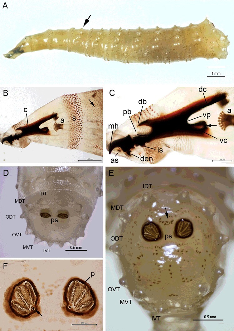
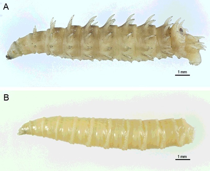
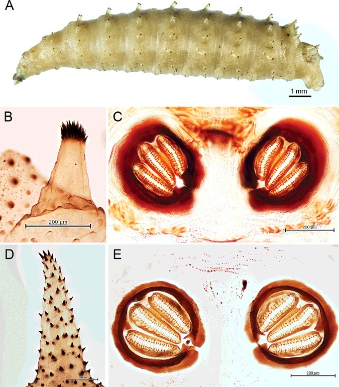
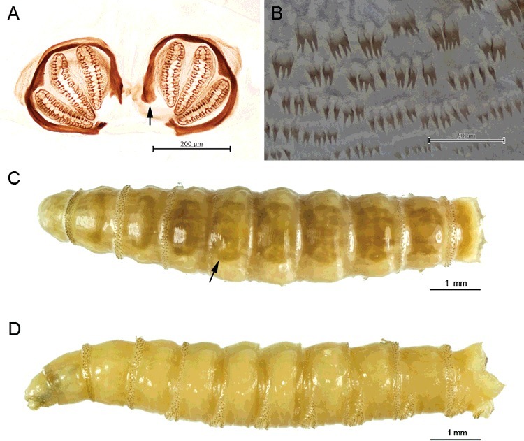
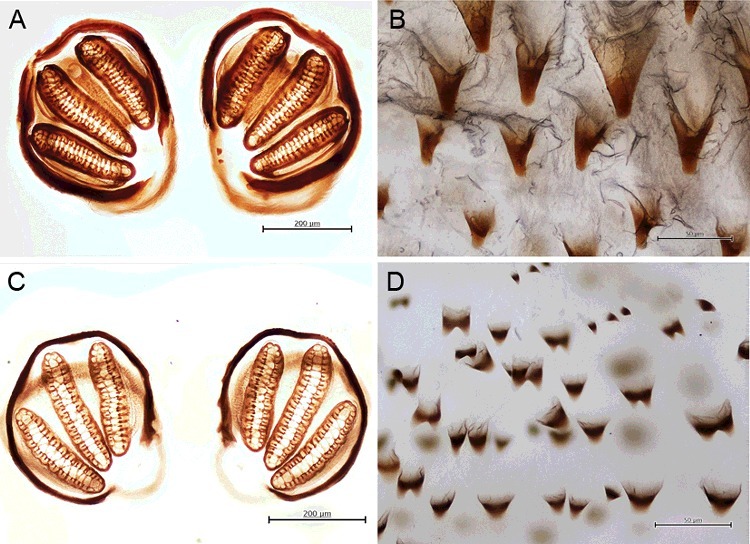
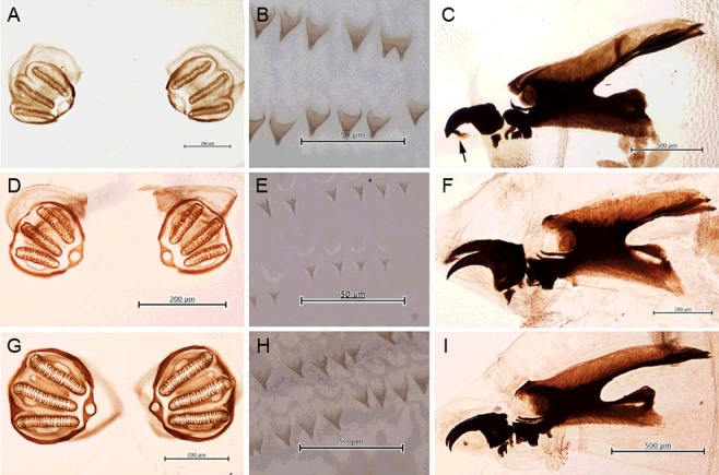
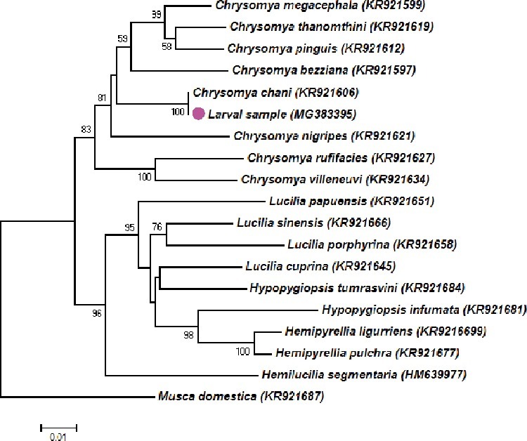
Blow flies are worldwide the most important insects from a forensic point of view. In Thailand, aside from the two most common species, Chrysomya megacephala (F.) and Chrysomya rufifacies (Macquart), Chrysomya chani Kurahashi was also found to be of forensic importance. We present a case of a human female cadaver in its bloated stage of decomposition, discovered at Pachangnoi Subdistrict, northern Thailand. Entomological sampling during the autopsy displayed an assemblage of numerous dipteran larvae. Macroscopic observations showed the coexistence of third instar larvae of the three blow flies C. megacephala, Chrysomya villeneuvi Patton, an unknown blow fly species and one muscid, Hydrotaea sp. The minimum post-mortem interval was estimated to be six days, based on the developmental rate of C. megacephala. The ID of the unknown larva, which is the focus of this report, was revealed later as C. chani by DNA sequencing, using a 1205 bp of cytochrome c oxidase subunit I (COI). The occurrence of C. chani on a human body revealed the need to analyse and describe the morphology of its immature stage, to enable forensic entomologists to identify this fly species in future cases. The morphological examination of the third instar was performed, revealing peculiar characteristics: protuberant tubercles encircling abdominal segments; 9-11 lobes on the anterior spiracle; six prominent pairs of tubercles along the peripheral rim of the eighth abdominal segment; a heavily sclerotized complete peritreme of the posterior spiracles. A key to differentiate the third instar of blow flies of forensic importance in Thailand is provided.
Keywords: Chrysomya chani; Forensic science; Thailand; forensic entomology; larval morphology, identification.
Figures


Similar articles
-
Geometry of posterior larval spiracles to identify medically and forensically important calliphorids in Thailand.Acta Trop. 2024 Apr;252:107126. doi: 10.1016/j.actatropica.2024.107126. Epub 2024 Feb 3. Acta Trop. 2024. PMID: 38316241
-
Ultrastructure of male genitalia of blow flies (Diptera: Calliphoridae) of forensic importance.Acta Trop. 2018 Mar;179:61-80. doi: 10.1016/j.actatropica.2017.12.014. Epub 2017 Dec 14. Acta Trop. 2018. PMID: 29248415
-
Forensic entomology cases in Thailand: a review of cases from 2000 to 2006.Parasitol Res. 2007 Oct;101(5):1417-23. doi: 10.1007/s00436-007-0659-8. Epub 2007 Jul 24. Parasitol Res. 2007. PMID: 17647017 Review.
-
The Importance of Habitat in the Ecology of Decomposition on Rabbit Carcasses in Malaysia: Implications in Forensic Entomology.J Med Entomol. 2015 Jan;52(1):9-23. doi: 10.1093/jme/tju001. J Med Entomol. 2015. PMID: 26336275
-
Chrysomya pinguis (Walker) (Diptera: Calliphoridae), blow fly of forensic importance: A review of bionomics and forensic entomology appraisal.Acta Trop. 2022 Aug;232:106506. doi: 10.1016/j.actatropica.2022.106506. Epub 2022 May 11. Acta Trop. 2022. PMID: 35562089 Review.
Cited by
-
Predicting Geographic Distribution of Forensically Significant Blow Flies of Subfamily Chrysomyinae (Diptera: Calliphoridae) in Northern Thailand.Insects. 2018 Aug 21;9(3):106. doi: 10.3390/insects9030106. Insects. 2018. PMID: 30134628 Free PMC article.
-
Origins and Diversification of Myiasis Across Blowflies.Ecol Evol. 2025 Feb 13;15(2):e70993. doi: 10.1002/ece3.70993. eCollection 2025 Feb. Ecol Evol. 2025. PMID: 39949886 Free PMC article.
-
Molecular Analysis of Forensically Important Blow Flies in Thailand.Insects. 2018 Nov 8;9(4):159. doi: 10.3390/insects9040159. Insects. 2018. PMID: 30413082 Free PMC article.
References
-
- Amendt J, Richards CS, Campobasso CP, et al. . Forensic entomology: applications and limitations. Forensic Sci Med Pathol. 2011;7:379–392. - PubMed
-
- Sukontason K, Sukontason KL, Piangjai S, et al. . Hairy maggot of Chrysomya villeneuvi (Diptera: Calliphoridae), a fly species of forensic importance. J Med Entomol. 2003;40:983–984. - PubMed
-
- Sukontason KL, Kanchai C, Piangjai S, et al. . Morphological observation of puparia of Chrysomya nigripes (Diptera: Calliphoridae) from human corpse. Forensic Sci Int. 2006;161:15–19. - PubMed
-
- Sukontason K, Narongchai P, Kanchai C, et al. . Forensic entomology cases in Thailand: a review of cases from 2000 to 2006. Parasitol Res. 2007;101:1417–1423. - PubMed
LinkOut - more resources
Full Text Sources
