. 2019 Jan;19(1):617-621.
doi: 10.3892/mmr.2018.9669. Epub 2018 Nov 19.

Magnetization transfer contrast MRI in GFP‑tagged live bacteria

Affiliations

Magnetization transfer contrast MRI in GFP‑tagged live bacteria

Valeria Righi et al. Mol Med Rep. 2019 Jan.

Abstract

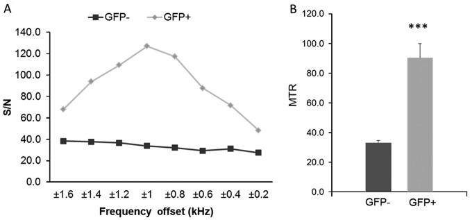
Green fluorescent protein (GFP) is a widely utilized molecular reporter of gene expression. However, its use in in vivo imaging has been restricted to transparent tissue mainly due to the tissue penetrance limitation of optical imaging. Magnetization transfer contrast (MTC) is a magnetic resonance imaging (MRI) methodology currently utilized to detect macromolecule changes such as decrease in myelin and increase in collagen content. MTC MRI imaging was performed to detect GFP in both in vitro cells and in an in vivo mouse model to determine if MTC imaging could be used to detect infection from Pseudomonas aeruginosa in murine tissues. It was demonstrated that the approach produces values that are protein specific and concentration dependent. This method provides a valuable, non‑invasive imaging tool to study the impact of novel antibacterial therapeutics on bacterial proliferation and perhaps viability within the host system, and could potentially suggest the modulation of bacterial gene expression within the host when exposed to such compounds.

Keywords: magnetization transfer contrast imaging; green fluorescent protein; Pseudomonas aeruginosa; burn mouse model; in vitro cells; in vivo mouse.


Figures

Figure 1.
Figure 1.
GFP-tagged Pseudomonas aeruginosa cells viewed at ×600 magnification. GFP, green fluorescent protein.
Figure 2.
Figure 2.
Pseudocolored pixel of EC-GFP, PA-GFP and PA at 0.25 kHz offset. GFP, green fluorescent protein; EC-GFP, GFP-tagged E. coli; PA-GFP, GFP-tagged Pseudomonas aeruginosa.
Figure 3.
Figure 3.
(A) Region based MTR calculations for the different frequency offsets for PA, PA-GFP and EC-GFP from 0.05 to 0.45 kHz (frequency offset). (B) MTR (mean ± SE) of PA, PA-GFP and EC-GFP. Data were analyzed using one-way ANOVA analysis followed by Tukey's post hoc test. **P<0.01 and ***P<0.001 vs. PA. PA, Pseudomonas aeruginosa; GFP, green fluorescent protein; EC-GFP, GFP-tagged E. coli; PA-GFP, GFP-tagged Pseudomonas aeruginosa; MTR, magnetization transfer ratio.
Figure 4.
Figure 4.
Pseudocolored pixel of GFP+ (upper) and GFP (lower) mice MR images at 1 kHz offset. GFP, green fluorescent protein.
Figure 5.
Figure 5.
(A) Region based MTR calculations for the different frequency offsets for mice GFP and mice GFP+ from 0.2 to 1.6 kHz (frequency offset). (B) MTR (mean ± SE) of GFP and GFP+, P-values. ***P<0.0001 vs. GFP. GFP, green fluorescent protein; MTR, magnetization transfer ratio.

Similar articles

References

    1. Turner KH, Everett J, Trivedi U, Rumbaugh KP, Whiteley M. Requirements for Pseudomonas aeruginosa acute burn and chronic surgical wound infection. PLoS Genet. 2014;10:e1004518. doi: 10.1371/journal.pgen.1004518. - DOI - PMC - PubMed
    1. Pendleton JN, Gorman SP, Gilmore BF. Clinical relevance of the ESKAPE pathogens. Expert Rev Anti Infect Ther. 2013;11:297–308. doi: 10.1586/eri.13.12. - DOI - PubMed
    1. Church D, Elsayed S, Reid O, Winston B, Lindsay R. Burn wound infections. Clin Microbiol Rev. 2006;19:403–434. doi: 10.1128/CMR.19.2.403-434.2006. - DOI - PMC - PubMed
    1. Lyczak JB, Cannon CL, Pier GB. Establishment of Pseudomonas aeruginosa infection: Lessons from a versatile opportunist. Microbes Infect. 2000;2:1051–1060. doi: 10.1016/S1286-4579(00)01259-4. - DOI - PubMed
    1. McVay CS, Velásquez M, Fralick JA. Phage therapy of Pseudomonas aeruginosa infection in a mouse burn wound model. Antimicrob Agents Chemother. 2007;51:1934–1938. doi: 10.1128/AAC.01028-06. - DOI - PMC - PubMed

MeSH terms