. 2019 Feb;54(2):443-454.
doi: 10.3892/ijo.2018.4647. Epub 2018 Nov 26.

MicroRNA-30e inhibits adhesion, migration, invasion and cell cycle progression of prostate cancer cells via inhibition of the activation of the MAPK signaling pathway by downregulating CHRM3

Affiliations

MicroRNA-30e inhibits adhesion, migration, invasion and cell cycle progression of prostate cancer cells via inhibition of the activation of the MAPK signaling pathway by downregulating CHRM3

Xin-Min Zheng et al. Int J Oncol. 2019 Feb.

Retraction in

Abstract

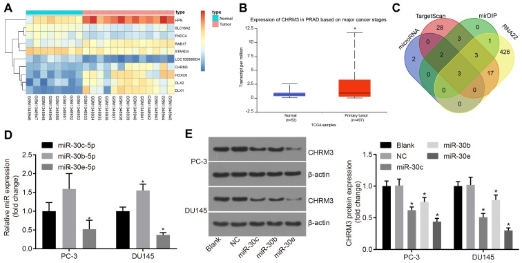
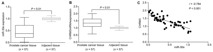
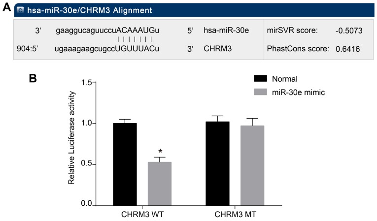
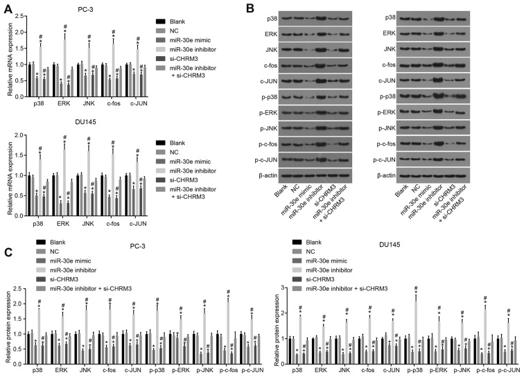
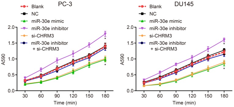
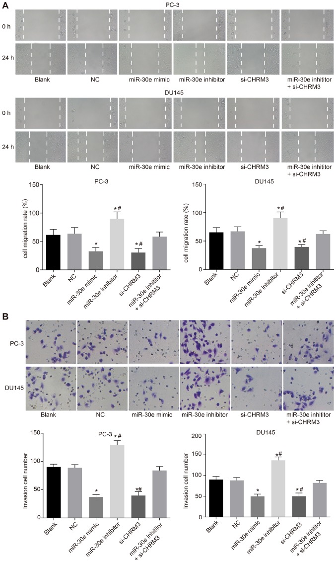
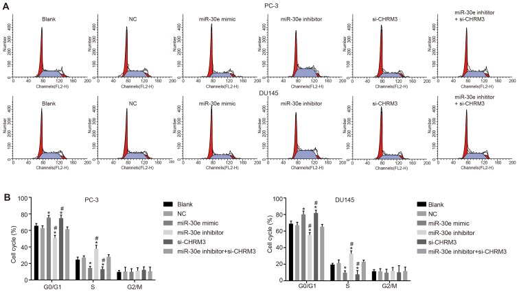
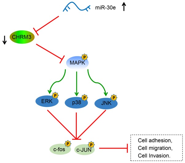
Prostate cancer (PCa) testing is currently based on measurement of serum prostate‑specific antigen levels and digital rectal examination, which are limited by a low predictive value and the adverse effects associated with overdiagnosis and overtreatment. Recent studies have reported that the abnormal expression of microRNAs (miRNAs) is associated with the mechanism underlying the development of PCa. Thus, the aim of the present study was to investigate the effects of miR‑30e and its target gene, M3 muscarinic acetylcholine receptor (CHRM3), on the adhesion, migration, invasion and cell cycle distribution of PCa cells via the mitogen‑activated protein kinase (MAPK) signaling pathway. The differentially expressed genes were screened in the Gene Expression Omnibus database from a gene expression microarray (GSE55945) of PCa. PCa tissues and adjacent tissues were collected from patients with PCa. The PC‑3 and DU145 human PCa cell lines were treated with activator, inhibitor and siRNAs. The effects of miR‑30e on cell adhesion, migration, invasion and cell cycle distribution with the involvement of CHRM3 and the MAPK signaling pathway were investigated. The bioinformatics results demonstrated that the CHRM3 gene and the MAKP signaling pathway were involved in the progression of PCa, and has‑miR‑30e was selected for further study. The levels of miR‑30e were significantly downregulated, while the levels of CHRM3 were obviously upregulated in PCa. CHRM3 was verified as a target gene of miR‑30e. Upregulation of miR‑30e and downregulation of CHRM3 decreased the levels of p‑P38, p‑extracellular signal‑regulated kinase, p‑c‑Jun N‑terminal kinase, p‑c‑fos and p‑c‑JUN, cell adhesion, migration and invasion ability, and the number of cells in the S phase, while they increased the number of cells in the G0 and G1 phases. The findings of the present study suggest that miR‑30e inhibited the adhesion, migration, invasion and cell cycle entry of PCa cells by suppressing the activation of the MAPK signaling pathway and inhibiting CHRM3 expression. Thus, miR‑30e may serve as a candidate target for the treatment of PCa.

Keywords: microRNA-30e; M3 muscarinic acetylcholine receptor; mitogen-activated protein kinase signaling pathway; prostate cancer cells; adhesion; migration; invasion; cell cycle.


Figures

Figure 1
Figure 1
Analysis of levels of CHRM3 and regulatory miRNAs of CHRM3 in PCa. (A) Differential analysis of GSE55945 chip data; the transverse coordinates represent the number of samples, an expression heat map of the differentially expressed genes was established, and the ordinate indicates the names of the differentially expressed genes. The upper right histogram represents color; from top to bottom, the color change indicates changes of expression in microarray data from large to small. Each rectangle in the graph corresponds to the expression value of a sample. Each column represents the expression of all the genes in each sample. The tree diagram on the left shows the results of cluster analysis of different genes from different samples, the top bars represent sample type, blue representing normal control samples, and red representing prostate cancer samples. (B) analysis of levels of CHRM3 in PCa in TCGA database exhibiting high levels of CHRM3. The left blue box graph represents the expression of CHRM3 in the 52 normal samples of TCGA database. The red box on the right shows the expression of CHRM3 in the 497 prostate cancer samples in TCGA database; *P<0.001. (C) has-miR-30c-5p, has-miR-30b-5p and has-miR-30e-5p appeared in the prediction results of four databases. The intersection of the results of the four databases was taken; blue represents prediction result of regulatory miRNAs of CHRM3 in microRNA.org, red represents prediction result of regulatory miRNAs of CHRM3 in TargetScan, green represents prediction results of regulatory miRNAs of CHRM3 in mirDIP, and yellow represents prediction result of regulatory miRNAs of CHRM3 in the RNA22 database. The blue arrow is the intersection of four database prediction results. (D) RT-qPCR was performed to detect the levels of has-miR-30c-5p, has-miR-30b-5p and has-miR-30e-5p in the PCa cell lines PC-3 and DU145, and the results demonstrated that the miR-30e-5p expression was the lowest in the PC-3 and DU145 cell lines. (E) Western blot analysis was performed to investigate the levels of has-miR-30c-5p, has-miR-30b-5p, has-miR-30e-5p and CHRM3, and the results demonstrated that miR-30e inhibited the expression of CHRM3 in PCa cells; *P<0.05; miR-30e, microRNA-30e; CHRM3, M3 muscarinic acetylcholine receptor; RT-qPCR, reverse transcription-quantitative polymerase chain reaction; PCa, prostate cancer; TCGA, The Cancer Genome Atlas.
Figure 2
Figure 2
RT-qPCR and western blot analysis revealed low levels of miR-30e and high levels of CHRM3 in PCa tissues, and miR-30e was negatively correlated with the mRNA levels of CHRM3. (A) The results of RT-qPCR demonstrated lower levels of miR-30e in PCa tissues compared with the adjacent tissues. (B) The results of RT-qPCR demonstrated higher mRNA levels of CHRM3 in PCa tissues compared with the adjacent tissues. (C) Pearson’s correlation analysis showed that miR-30e was negatively correlated with the mRNA levels of CHRM3 in PCa tissues; n=57; P<0.01, P<0.001 were considered significant; RT-qPCR, reverse transcription-quantitative polymerase chain reaction; miR-30e, microRNA-30e; CHRM3, M3 muscarinic acetylcholine receptor; PCa, prostate cancer.
Figure 3
Figure 3
Biological information prediction and double luciferase reporter gene assays revealed that CHRM3 is a target gene of miR-30e. (A) miR-30e combined with CHRM3-3′-UTR predicted by a public database. (B) Dual-luciferase reporter gene assays revealed that CHRM3 is a potential target gene of miR-30e; *P<0.001 compared with WT + mimic. WT, wild-type; MT, mutant; UTR, untranslated region; miR-30e, microRNA-30e; CHRM3, M3 muscarinic acetylcholine receptor; PCa, prostate cancer.
Figure 4
Figure 4
The results of RT-qPCR and western blot analysis revealed that miR-30e inhibits the expression of CHRM3 in PCa cells. (A) The mRNA level of miR-30e in the miR-30e mimic and si-CHRM3 groups was downregulated. (B) The protein level of CHRM3 in the miR-30e inhibitor + si-CHRM3 group was obviously increased. Blank group, no plasmids were transfected; negative control (NC) group, transfected with 50 nM negative and nonsense sequence; miR-30e mimic group, transfected with 50 nM miR-30e mimic sequence; miR-30e inhibitor group, transfected with 50 nM miR-30e inhibitor sequence; si-CHRM3 group, transfected with 50 nM CHRM3-siRNA plasmid; miR-30e inhibitors + si-CHRM3 group, transfected with 50 nM miR-30e inhibitor sequence and 50 nM CHRM3-siRNA plasmid. *P<0.05 compared with the blank and NC groups; #P<0.05 compared with the si-CHRM3 + miR-30e inhibitor group. RT-qPCR, reverse transcription-quantitative polymerase chain reaction; miR-30e, microRNA-30e; CHRM3, M3 muscarinic acetylcholine receptor; PCa, prostate cancer.
Figure 5
Figure 5
Upregulation of miR-30e lowers the levels of the MAPK signaling pathway genes (p38, ERK and JUN) and downstream genes (c-fos and c-JUN) via CHRM3 in PCa cells as shown by RT-qPCR and western blot analysis. (A) Τhe miR-30e inhibitor group had increased levels of the signaling pathway genes (p38, ERK and JUN) and downstream genes (c-fos and c-JUN) of the MAPK signaling pathway. (B) The si-CHRM3 group exhibited reduced levels of p38, ERK, JUN, c-fos, c-JUN and phosphorylated proteins (p-P38, p-ERK, p-JNK, p-c-fos and p-c-JUN) of the MAPK signaling pathway. (C) Gray values of the related proteins oν western blot analysis; blank group, no plasmids were transfected; negative control (NC) group, transfected with 50 nM negative and nonsense sequence; miR-30e mimic group, transfected with 50 nM miR-30e mimic sequence; miR-30e inhibitor group, transfected with 50 nM miR-30e inhibitor sequence; si-CHRM3 group, transfected with 50 nM CHRM3-siRNA plasmid; miR-30e inhibitors + si-CHRM3 group, transfected with 50 nM miR-30e inhibitor sequence and 50 nM CHRM3-siRNA plasmid. *P<0.05 compared with the blank and NC groups; #P<0.05 compared with the si-CHRM3 + miR-30e inhibitor group. RT-qPCR, reverse transcription-quantitative polymerase chain reaction; miR-30e, microRNA-30e; CHRM3, M3 muscarinic acetylcholine receptor; PCa, prostate cancer; ERK, extracellular signal-regulated kinase; JNK, c-Jun N terminal kinase.
Figure 6
Figure 6
Upregulation of miR-30e is involved in the inhibition of the adhesion process of DU145 and PC-3 cells as determined by MTT assays. Blank group, no plasmids were transfected; negative control (NC) group, transfected with 50 nM negative and nonsense sequence; miR-30e mimic group, transfected with 50 nM miR-30e mimic sequence; miR-30e inhibitor group, transfected with 50 nM miR-30e inhibitor sequence; si-CHRM3 group, transfected with 50 nM CHRM3-siRNA plasmid; miR-30e inhibitors + si-CHRM3 group, transfected with 50 nM miR-30e inhibitor sequence and 50 nM CHRM3-siRNA plasmid. MTT, 3-(4,5-dimethylthiazol-2-yl)-2,5-diphenyltetrazolium bromide; miR-30e, microRNA-30e; CHRM3, M3 muscarinic acetylcholine receptor; ERK, extracellular signal-regulated kinase; MAPK, mitogen-activated protein kinase; JNK, c-Jun N terminal kinase; PCa, prostate cancer.
Figure 7
Figure 7
Upregulation of miR-30e inhibits migration and invasion of PCa cells as verified by scratch tests and Transwell assays. (A) Upregulation of miR-30e inhibits the migration of DU145 and PC-3 cells as shown by the scratch test. (B) Upregulation of miR-30e inhibits invasion of DU145 and PC-3 cells as shown by Transwell assays; *P<0.05 compared with the blank and negative control (NC) groups; #P<0.05 compared with the si-CHRM3 + miR-30e inhibitor group. miR-30e, microRNA-30e; CHRM3, M3 muscarinic acetylcholine receptor; PCa, prostate cancer.
Figure 8
Figure 8
Upregulation of miR-30e inhibits cell cycle progression of DU145 and PC-3 cells as shown by PI single staining. (A) The number of cells in the G1 phase obviously increased and the number of cells in the S phase markedly decreased in the miR-30e mimic and si-CHRM3 groups, while the results in the miR-30e inhibitor group were the opposite. (B) Upregulation of miR-30e decreased the number of cells in the S phase, while it increased the number of cells in the G0 and G1 phases. *P<0.05 compared with the blank and negative control (NC) groups; miR-30e, microRNA-30e; CHRM3, M3 muscarinic acetylcholine receptor; PCa, prostate cancer.
Figure 9
Figure 9
miR-30e inhibits adhesion, migration, invasion and cell cycle progression of PCa cells via suppressing CHRM3 expression and activation of the MAPK signaling pathway. The miR-30e mimic inhibited the expression of the CHRM3 gene and inhibited the activation of the MAPK signaling pathway, including the phosphorylation of ERK and JNK, and inhibited the activation of downstream c-fos and JUN, thereby inhibiting the adhesion, migration and invasion of PCa cells. CHRM3, M3 muscarinic acetylcholine receptor; PCa, prostate cancer; MAPK, mitogen-activated protein kinase; ERK extracellular signal-regulated kinase; JNK, c-Jun N terminal kinase.

Similar articles

Cited by

References

    1. Quinn DI, Shore ND, Egawa S, Gerritsen WR, Fizazi K. Immunotherapy for castration-resistant prostate cancer: Progress and new paradigms. Urol Oncol. 2015;33:245–260. doi: 10.1016/j.urolonc.2014.10.009. - DOI - PubMed
    1. Taylor BS, Schultz N, Hieronymus H, Gopalan A, Xiao Y, Carver BS, Arora VK, Kaushik P, Cerami E, Reva B, et al. Integrative genomic profiling of human prostate cancer. Cancer Cell. 2010;18:11–22. doi: 10.1016/j.ccr.2010.05.026. - DOI - PMC - PubMed
    1. Mahn R, Heukamp LC, Rogenhofer S, von Ruecker A, Muller SC, Ellinger J. Circulating microRNAs (miRNA) in serum of patients with prostate cancer. Urology. 2011;77(1265):e1269–1216. doi: 10.1016/j.urology.2011.01.020. - DOI - PubMed
    1. Wen X, Deng FM, Wang J. MicroRNAs as predictive biomarkers and therapeutic targets in prostate cancer. Am J Clin Exp Urol. 2014;2:219–230. - PMC - PubMed
    1. Lynch SM, McKenna MM, Walsh CP, McKenna DJ. miR-24 regulates CDKN1B/p27 expression in prostate cancer. Prostate. 2016;76:637–648. doi: 10.1002/pros.23156. - DOI - PubMed

Publication types

Substances