. 2019 Feb;54(2):505-514.
doi: 10.3892/ijo.2018.4638. Epub 2018 Nov 19.

Mitogen-activated kinase kinase kinase 1 inhibits hedgehog signaling and medulloblastoma growth through GLI1 phosphorylation

Affiliations

Mitogen-activated kinase kinase kinase 1 inhibits hedgehog signaling and medulloblastoma growth through GLI1 phosphorylation

Laura Antonucci et al. Int J Oncol. 2019 Feb.

Abstract

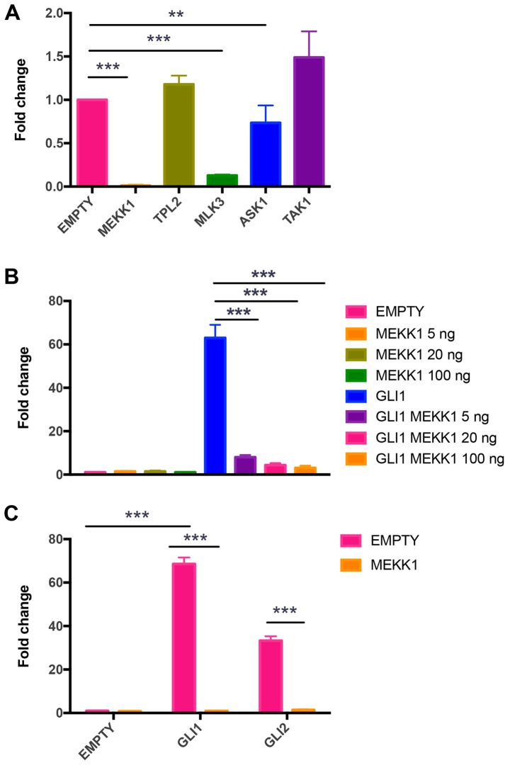
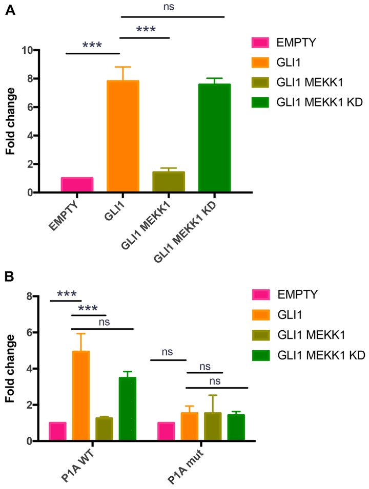
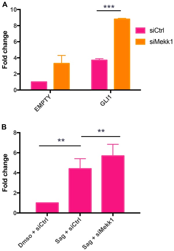
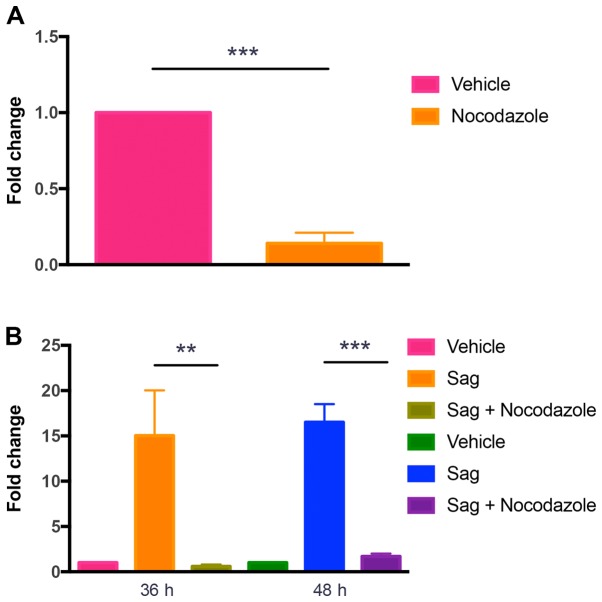
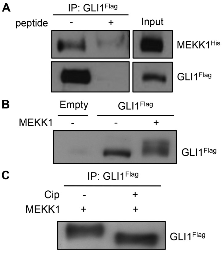
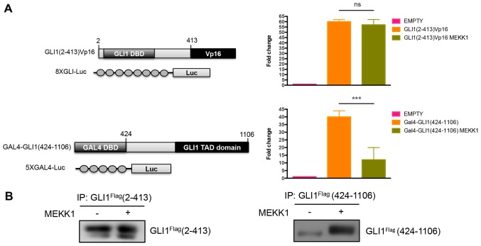
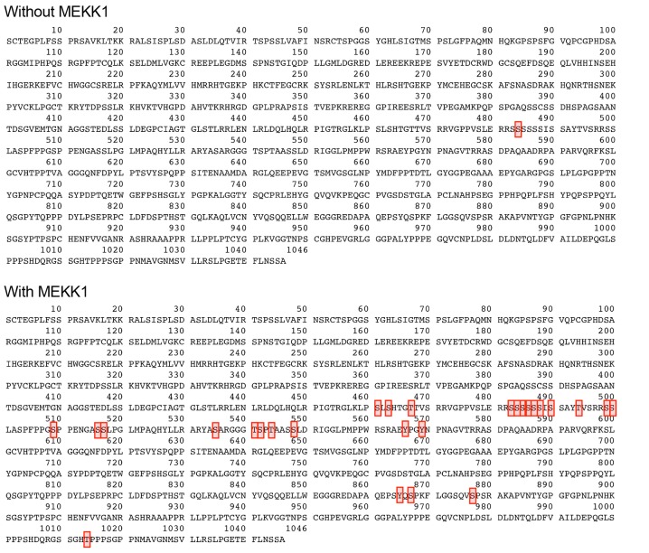
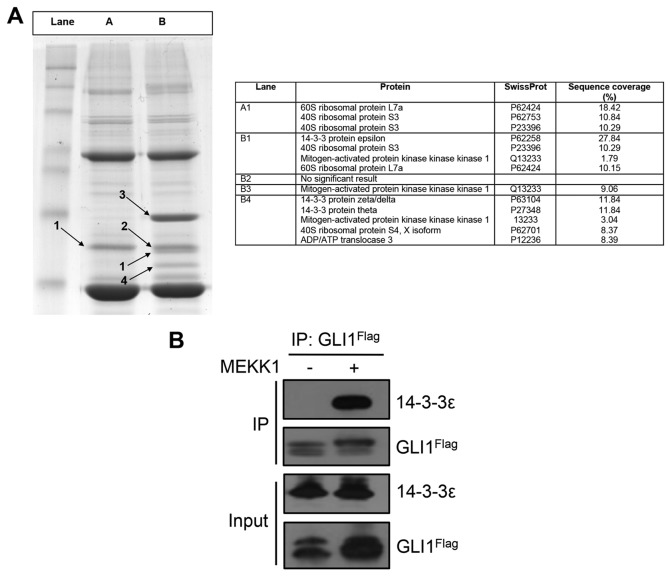
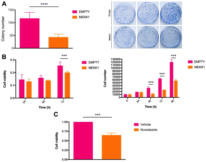
The aberrant activation of hedgehog (HH) signaling is a leading cause of the development of medulloblastoma, a pediatric tumor of the cerebellum. The FDA‑approved HH inhibitor, Vismodegib, which targets the transmembrane transducer SMO, has shown limited efficacy in patients with medulloblastoma, due to compensatory mechanisms that maintain an active HH‑GLI signaling status. Thus, the identification of novel actionable mechanisms, directly affecting the activity of the HH‑regulated GLI transcription factors is an important goal for these malignancies. In this study, using gene expression and reporter assays, combined with biochemical and cellular analyses, we demonstrate that mitogen‑activated kinase kinase kinase 1 (MEKK1), the most upstream kinase of the mitogen‑activated protein kinase (MAPK) phosphorylation modules, suppresses HH signaling by associating and phosphorylating GLI1, the most potent HH‑regulated transcription factor. Phosphorylation occurred at multiple residues in the C‑terminal region of GLI1 and was followed by an increased association with the cytoplasmic proteins 14‑3‑3. Of note, the enforced expression of MEKK1 or the exposure of medulloblastoma cells to the MEKK1 activator, Nocodazole, resulted in a marked inhibitory effect on GLI1 activity and tumor cell proliferation and viability. Taken together, the results of this study shed light on a novel regulatory mechanism of HH signaling, with potentially relevant implications in cancer therapy.

Keywords: hedgehog; medulloblastoma; GLI1; mitogen-activated kinase kinase kinase 1; phosphorylation.


Figures

Figure 1
Figure 1
Regulation of the GLI1 promoter by MEKK1. (A) 293T cells were co-transfected with the expression plasmid encoding Flag-GLI1 and expression vectors for MEKK1, TPL2, MLK3, ASK1, TAK1 or control vector (Flag-pcDNA3), and a 12x-GLI luciferase promoter reporter. Plasmid TK-Renilla was transfected to normalize the transfection efficiency. Data are expressed as the fold change of the ratio between luciferase to Renilla activity. (B) 293T cells were transfected with 8x-GLI luciferase promoter reporter and TK Renilla plasmids and with vectors expressing Flag-pcDNA3 or Flag-GLI1 and increasing amounts of MEKK1 plasmid (5, 20 and 100 ng), where indicated. Data are shown as the fold change of the ratio of luciferase reporter to Renilla activity. (C) 293T cells were co-transfected with 8x-GLI luciferase reporter and TK Renilla plasmids and Flag-GLI1 or Flag-GLI2 expression vectors or control vector (Flag-pcDNA3). Data are expressed as the fold change of the ratio between luciferase to Renilla activity. Results are the means ± SD of 3 independent experiments, each performed in triplicate. **P<0.01 and ***P<0.001 (determined by ANOVA with Tukey’s post hoc test).
Figure 2
Figure 2
MEKK1 inhibits promoter-specific PTCH1 transcription. (A) NIH3T3 cells were transfected with Flag-GLI1 or control vector and MEKK1 expression plasmids. Relative expression of PTCH1 mRNA was evaluated by RT-qPCR. Data were normalized to HPRT mRNA levels and are expressed as the fold change relative to control sample. (B) 293T cells were transiently transfected with P1A WT and P1A mutant promoter luciferase constructs, and the TK Renilla encoding gene as reporters, and plasmids encoding GLI1, MEKK1 WT or KD or empty vector. Data are expressed as the fold change of the ratio between luciferase to Renilla activity. The results are the means ± SD of 3 independent experiments, each performed in triplicate. ***P<0.001; ns, not significant (calculated by ANOVA with Tukey’s post hoc test).
Figure 3
Figure 3
MEKK1 ablation increases HH/GLI1 activity. (A) NIH3T3 cells were transiently transfected with control siRNA (siCtrl, 100 nM) or Mekk1 siRNA (siMekk1, 100 nM) and 12x-GLI and TK Renilla luciferase reporters. Cells were harvested and analyzed for luciferase activity. Data are expressed as the fold change of the ratio between luciferase to Renilla activity. (B) GLI1 mRNA expression levels in Sag-treated NIH3T3 cells expressing control siRNA (siCtrl, 100 nM) or Mekk1 siRNA (siMekk1, 100 nM) were evaluated by RT-qPCR. GLI1 mRNA levels were normalized to HPRT mRNA levels and expressed as fold change relative to control sample. Data are the means ± SD of at least 3 independent experiments, each performed in triplicate. **P<0.01 and ***P<0.001 (determined by ANOVA with Tukey’s post hoc test).
Figure 4
Figure 4
Pharmacological MEKK1 activation inhibits GLI1 activity. (A) Luciferase reporter activity in 293T cells transiently transfected with 12x-GLI luciferase and TK Renilla reporters. Cells were treated with Nocodazole (1 µg/ml) for 24 h and analyzed for luciferase activity. Data are expressed as the fold change of the ratio between luciferase to Renilla activity. (B) GLI1 mRNA expression levels in Sag-treated NIH3T3 cells exposed to Nocodazole (0.1 µg/ml) for 36 and 48 h. GLI1 were analyzed by RT-qPCR and normalized to GAPDH mRNA levels. Results are expressed as the fold change relative to the control (vehicle) sample. Data are the means ± SD of at least 3 independent experiments, each performed in triplicate. **P<0.01 and ***P<0.001 [calculated using the Student’s t-test (A) or ANOVA with Tukey’s post hoc test (B)].
Figure 5
Figure 5
MEKK1 binds and phosphorylates GLI1. (A) 293T cells were trans-fected with constructs encoding Flag-GLI1 and His-MEKK1 and cell lysates were immunoprecipitated with anti-Flag antibody without or with 0.1 mg/ml blocking peptide. MEKK1 and GLI1 were revealed with anti MEKK1 and anti-Flag antibodies, respectively. Input: 5%. (B) Western blot analysis of cellular lysates from 293T cells expressing Flag-Gli1 alone or in combination with His-MEKK1. Flag-GLI1 was revealed with anti-Flag antibody. (C) 293T cells transfected with plasmids encoding Flag-GLI1 and His-MEKK1. Cell lysates were immunoprecipitated and immunocomplexes were incubated in the absence or presence of calf intestinal phosphatase (CIP).
Figure 6
Figure 6
MEKK1 inhibits GLI1 transcriptional activity and phosphorylates the C-terminal region of GLI1. (A) Left panel, schematic representation of fusion vectors expressing GLI1(2-413)Vp16 and 8xGLI luciferase reporter or Gal4-GLI1(424-1106) and 5xGal4 luciferase reporter plasmids. Right panel, luciferase assay on 293T cells transfected with GLI1(2-413)Vp16 and 8xGli-Luc or Gal4-GLI1(424-1106) and 5xGal4-Luc vectors, with or without MEKK1 expression plasmid. Plasmid encoding TK Renilla was transfected to normalize transfection efficiency. Data are expressed as the fold change of the ratio between luciferase to Renilla activity. (B) Western blot analysis of immunoprecipitates from 293T cells transfected with expression vectors for Flag-GLI1(2-413) or Flag-GLI1(424-1106), with or without MEKK1. Flag-GLI1 was revealed with anti-Flag antibody. Results are expressed as the means ± SD of at least 3 independent experiments, each performed in triplicate. ***P<0.001; ns, not significant (calculated by ANOVA with Tukey’s post hoc test).
Figure 7
Figure 7
Phosphorylation status of GLI1 in absence or presence of MEKK1. Aminoacidic sequence of GLI1 showing phosphorylated residues (highlighted in red). Mass spectrometric analysis was performed on 293T cells transfected with plasmid encoding Flag-GLI1 C-terminal region (AA 424-1106), with or without MEKK1. Cell lysates were immunoprecipitated with anti-Flag antibody and analyzed.
Figure 8
Figure 8
(A) Left panel, Coomassie-blue stained SDS-PAGE gel, showing proteins associated to Flag-GLI1(424-1106), in the absence or presence of MEKK1 in 293T cells. Lane A, proteins associated with Flag-GLI1(424-1106); lane B, proteins associated with Flag-GLI1(424-1106) + MEKK1. Numbered black arrows indicate bands cut out from the gel and processed. Right panel, mass spectrometry results for the selected gel bands. (B) Co-immunoprecipitation assay of Flag-GLI1 with endogenous 14-3-3ε proteins in 293T cells, with or without MEKK1. Lysates were immunoprecipitated with anti-Flag antibody and endogenous 14-3-3ε proteins binding is shown.
Figure 9
Figure 9
MEKK1 counteracts in vitro medulloblastoma cells growth and viability. (A) Colony formation assay on Med1-MB cells stably transfected with expression plasmids encoding MEKK1 or empty vector. Left panel, colonies were quantified with ImageJ software (Version 1.50i), using 4 images per dish. The bar graph represents the average of three biological replicates. Right, representative colony formation assay. (B) Left panel, MTT assay measuring cell proliferation activity of Med1-MB cells expressing MEKK1 or control plasmids at 24 h, 48 h and 72 h in each group. Right panel, growth curve on Med1-MB cells expressing MEKK1 or empty vector. (C) MTT assay on Med1-MB cells treated with Nocodazole (0.1 µg/ml) for 48 h. Results are expressed as the means ± SD of 3 independent experiments, each performed in triplicate. ***P<0.001 and ****P<0.0001 [calculated by a Student’s t-test (A and C) or ANOVA with Tukey’s post hoc test (B)].

References

    1. Dellovade T, Romer JT, Curran T, Rubin LL. The hedgehog pathway and neurological disorders. Annu Rev Neurosci. 2006;29:539–563. doi: 10.1146/annurev.neuro.29.051605.112858. - DOI - PubMed
    1. Ryan KE, Chiang C. Hedgehog secretion and signal transduction in vertebrates. J Biol Chem. 2012;287:17905–17913. doi: 10.1074/jbc.R112.356006. - DOI - PMC - PubMed
    1. Hui CC, Angers S. Gli proteins in development and disease. Annu Rev Cell Dev Biol. 2011;27:513–537. doi: 10.1146/annurev-cellbio-092910-154048. - DOI - PubMed
    1. Northcott PA, Jones DT, Kool M, Robinson GW, Gilbertson RJ, Cho YJ, Pomeroy SL, Korshunov A, Lichter P, Taylor MD, et al. Medulloblastomics: The end of the beginning. Nat Rev Cancer. 2012;12:818–834. doi: 10.1038/nrc3410. - DOI - PMC - PubMed
    1. Northcott PA, Korshunov A, Pfister SM, Taylor MD. The clinical implications of medulloblastoma subgroups. Nat Rev Neurol. 2012;8:340–351. doi: 10.1038/nrneurol.2012.78. - DOI - PubMed

MeSH terms