. 2019 Feb;97(2):177-188.
doi: 10.1007/s00109-018-1724-8. Epub 2018 Nov 27.

Interleukin-23 receptor signaling mediates cancer dormancy and radioresistance in human esophageal squamous carcinoma cells via the Wnt/Notch pathway

Affiliations

Interleukin-23 receptor signaling mediates cancer dormancy and radioresistance in human esophageal squamous carcinoma cells via the Wnt/Notch pathway

Yuepeng Zhou et al. J Mol Med (Berl). 2019 Feb.

Abstract

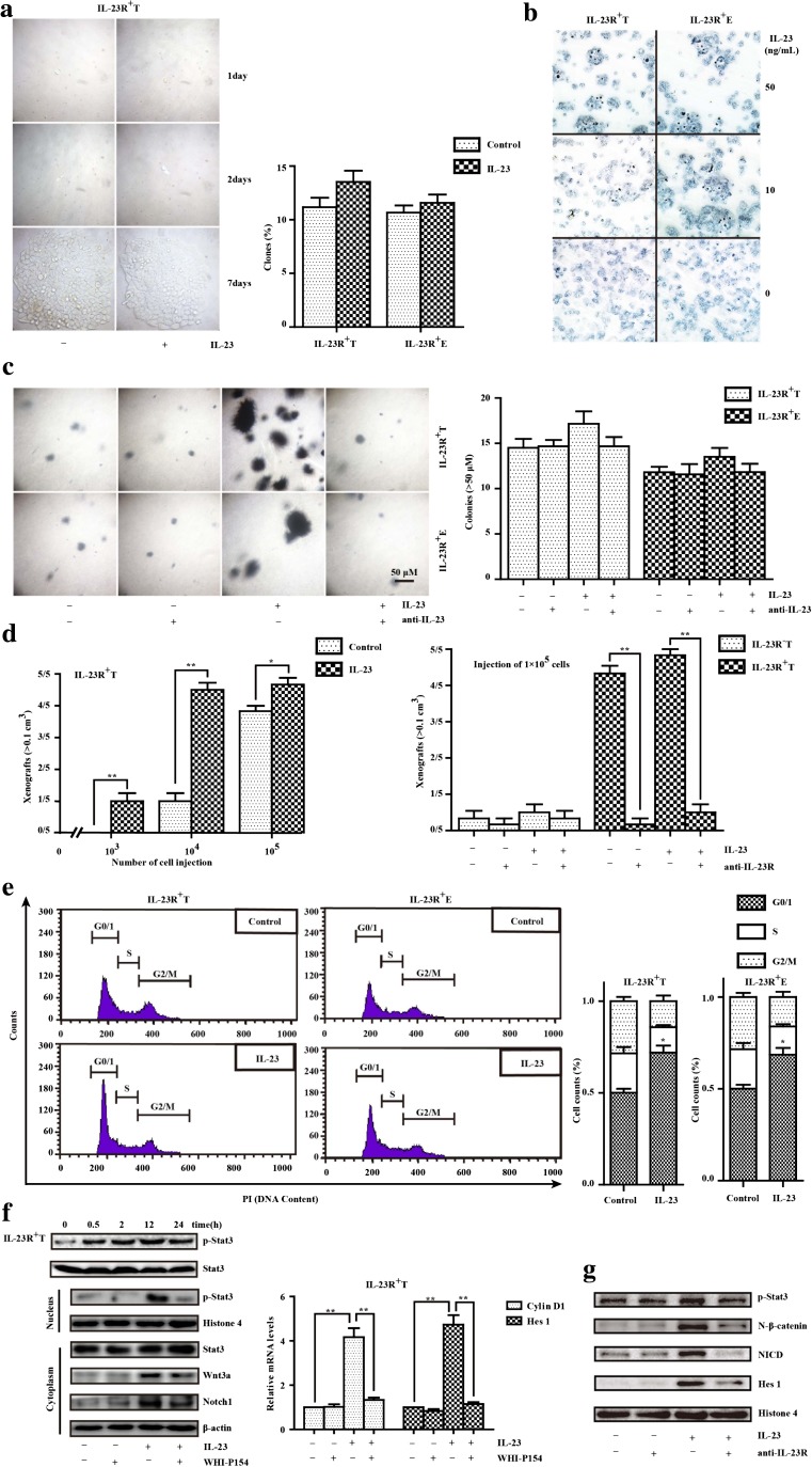
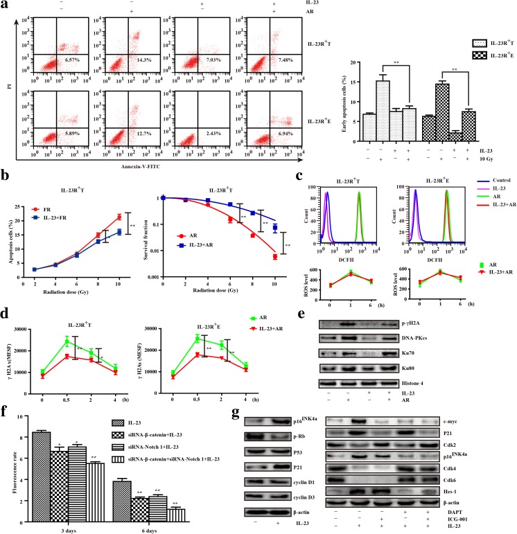
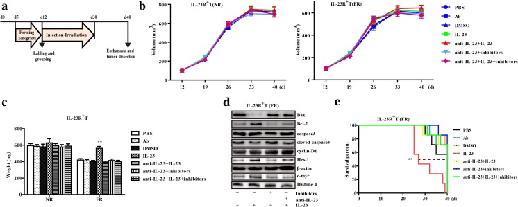
In the tumor microenvironment, inflammatory cells and molecules influence almost every process; among them, interleukin-23 (IL-23) is a pro-inflammatory molecule that exhibits pro- or anti-tumor properties, but both activities remain poorly understood. In this study, we investigated the effect of extracellular IL-23 in IL-23 receptor-positive (IL-23R+) esophageal squamous cell carcinoma (ESCC) and explored the mechanisms underlying this effect. We analyzed ESCC tumor tissues by immunohistochemical and immunofluorescence staining and found that IL-23, which was highly expressed, co-localized with Oct-4A in IL-23R+ ESCC cells. In addition, IL-23 treatment significantly increased the accumulation of CD133+ cells and activated the Wnt and Notch signaling pathways in CD133-IL-23R+ ESCC cell lines. Consistently, CD133-IL-23R+ cells pretreated with IL-23 showed stronger anti-apoptosis activity when exposed to radiation and higher survival than untreated groups. Moreover, the inhibition of Wnt/Notch signaling by a small-molecule inhibitor or siRNA abolished the effect of IL-23-induced dormancy and consequent radioresistance. Taken together, these results suggested that IL-23 facilitates radioresistance in ESCC by activating Wnt/Notch-mediated G0/1 phase arrest, and attenuating these detrimental changes by blocking the formation of dormancy may prove to be an effective pretreatment for radiotherapy. KEY MESSAGES: IL-23/IL-23R is correlated with the acquisition of stem-like potential in ESCC. CD133-IL-23R+ ESCCs acquired dormancy via IL-23. Radioresistance depends on IL-23-mediated Wnt/Notch pathway activation in vitro and vivo.

Keywords: Biomarker; Interleukin-23; M1 macrophage; Radioresistance; Wnt/notch.


Conflict of interest statement

Conflict of interest

The authors declare that they have no conflicts of interest.

Figures

Fig. 1
Fig. 1
The correlation between IL-23/IL-23R and the trans-differentiation of ESCCs. a Typical immunofluorescence images of the distribution of Oct-4A+ cells (red) and IL-23R+ ESCCs (green) gathering with the intensity of IL-23 (IHC). Left panel, × 200 magnification. Middle and right panels are magnifications of the area marked by dashed lines. b Western blotting: the expression of CD133 in ESCC cells (TE-1, ECA 109, KYSE 150, and TE-10) and Het-1A cells with or without IL-23 treatment (50 ng/mL, 24 h). The results were normalized to β-actin as a control and densitometric analysis of bands was performed with Alpha View. T, TE-1 cells; E, ECA 109 cells; H, Het-1A cells. c The percentage of CD133+ cells in ESCCs and Het-1A cells before and after sorting. Flow cytometry and fluorescent cell sorting were performed using anti-CD133 fluorescent-labeled antibody. The representative experiment results were compared with that untreated groups. d The number of protogenetic IL-23R+ cells. Flow cytometry was performed using anti-IL-23R (FITC) in ESCCs and Het-1A cells. The experiment was repeated twice and representative data shown. e The variants of CD133+ cells between IL-23R/IL-23R+ CD133ESCCs and Het-1A cells cultured with IL-23 (50 ng/mL, 24 h). f CD133IL-23R+ ESCCs and Het-1A cells were pretreated with IL-23 (50 ng/mL) for 24 h, the expression levels of CD133 were detected by Western blotting at 0, 24, 48, and 72 h after removing IL-23. g The relative mRNA expression levels of stemness genes (c-myc and Oct-4A) were measured by RT-PCR in CD133IL-23R+ ESCCs and Het-1A cells cultured with IL-23 (50 ng/mL) for 24 h. The GAPDH was used as the loading control. TE-1, ECA 109 cells, and Het-1A cells were treated with IL-23 for 48 h or not, and the protein expression of Oct-4A and c-myc was determined by Western blot analysis. β-Actin was used as a loading control. The data are presented as the mean ± SD from at least three independent experiments. **p < 0.01
Fig. 2
Fig. 2
The stem-like potencies of CD133IL-23R+ ESCCs stimulated by IL-23. a The plate clonogenic assay was performed in ESCCs. The CD133IL-23R+ TE-1 and ECA 109 cells were cultured with IL-23 (50 ng/mL) or not for 7 days. IL-23 had no significant impact on the clonogenicity ability of TE-1 cells (left panel). The statistical results (right panel) are the mean ± SEM (n = 3). b After the IL-23 treatment (10 or 50 ng/mL, 24 h) or not, CD133IL-23R+ TE-1 and ECA 109 cells expressing alkaline phosphatase (AKP) were counted. c The tumorigenic potentials with IL-23 treatment in CD133IL-23R+ TE-1 and ECA 109 cells were measured by the soft agar assay. Cells were cultured for 10 days in serum-free DMEM in the presence or absence of IL-23 (50 ng/mL) and/or IL-23 antibody (1 μg/mL). The results are the mean ± SEM (n = 3) (right panel). d The pretreatment of anti-IL-23R (0.5 μg) reversed the significant difference between the injection of IL-23 (50 ng) or vehicle groups that comprised IL-23R+ TE-1 cells (left panel). CD133IL-23R+ TE-1 cells were transplanted into nude mice in serial limiting dilutions (100,000, 10,000, or 1000 cells per injection) to form xenografts (> 0.1 cm3). e Phase of cell-cycle analysis by flow cytometry showed that IL-23R+ ESCC arrest at G0/1 correlated with IL-23 (50 ng/mL, 24 h) in CD133IL-23R+ TE-1 and ECA 109 cells. f The protein expressions of p-Stat3/Stat3, Wnt 3a, Notch 1, and the activation of the Wnt and Notch pathways (Cyclin D1 and Hes 1) were assessed by Western blotting and RT-PCR. The Western blotting results were normalized to β-actin or Histone 4 as the control. Cells were pretreated with WHI-P154 (2 μM) 2 h before adding IL-23 (50 ng/mL). IL-23 treatment (50 ng/mL, 24 h) activated Wnt/Notch signaling and the persistent phosphorylation level of Stat3 was maintained up to 24 h in TE-1 cells (left panel). Relative mRNA level of Cyclin D1 and Hes 1 were detected by RT-PCR in TE-1 cells (right panel). Compiled data were produced from three independent experiments. **p < 0.01. g Western blotting assessed the proteins expression of Stat3, Wnt, and Notch pathway. Cells were pretreated with anti-IL-23R (1 μg/mL) 2 h before adding IL-23 (50 ng/mL). The Western blotting results were normalized to β-actin or Histone 4 as the control
Fig. 3
Fig. 3
The Wnt/Notch signaling-mediated cell-cycle arrest due to IL-23 resulted in radioresistance. a The representative percentage of early apoptotic CD133IL-23R+ TE-1 and ECA 109 cells evaluated by Annexin-V and PI uptake after exposure to 10 Gy of ablative radiation (AR) with or without IL-23 treatment (50 ng/mL, 24 h) (left panel). The results are the mean ± SEM (n = 3, **p < 0.01) (right panel). b Early apoptotic CD133IL-23R+ TE-1 cells after each exposure to FR (2 Gy per fraction up to 10 Gy) with or without IL-23 treatment (50 ng/mL, 48 h) (left panel); SF of colonies were counted after cells exposed to AR (0, 2, 4, 6, 8, 10 Gy) with or without IL-23 treatment (50 ng/mL, 24 h) (middle panel). c According to the DCFH-DA fluorescence level by flow cytometry, the evaluation of cellular ROS levels after the exposure of CD133IL-23R+ ESCCs to AR (0, 1, and 6 h) with or without IL-23 (50 ng/mL, 24 h). d Formation of γH2AX was visualized in CD133IL-23R+ ESCCs after 10 Gy of AR (0, 0.5, 2, and 4 h) or pretreatment with IL-23 (50 ng/mL, 24 h). e The expression of p-γH2A, DNA-PKcs, Ku70, and Ku80 were detected in CD133IL-23R+ TE-1 cells cultured with IL-23 (50 ng/mL, 24 h) or not which were exposed to 10 Gy of AR by Western blotting. f CD133IL-23R+ TE-1 cells were transduced with non-silencing control siRNA (data not shown), siRNA-β-catenin, and/or siRNA-Notch1before the treatment of IL-23 (50 ng/mL). Cell proliferation was assessed by flow cytometry for CFSE labeling on the third and sixth days. g The protein expression changes of the cell cycle–regulating factors and Wnt/Notch signaling-related proteins from the IL-23 treatment (50 ng/mL, 24 h) and/or inhibitors (DAPT at 2 μM; ICG-001 at 5 μM) were detected by Western blotting. Compiled data were produced from three independent experiments. **p < 0.01
Fig. 4
Fig. 4
The in vivo study of IL-23 and radioresistance in a xenograft mouse model of ESCC. a Graphical representation of the experimental protocol and treatment schedule for stimulation and radiation in mice. b Line chart represents the average periodical tumor volume every week. The growth curves of CD133IL-23R+TE-1 cell xenograft tumors receiving different treatments, including PBS, isotype Ab, DMSO vehicle, irradiation, anti-IL-23 (1 μg in 100 μL PBS), human recombinant IL-23 (50 ng in 100 μL PBS), and the composite inhibitors (DAPT, 20 μmol and ICG-001, 50 μmol dissolved with DMSO in 100 μL PBS per day). The tumor volume was calculated as described in the “Materials and methods” section. NR, no-irradiation treatment; FR, fractionated radiation. c Column chart of xenograft tumor weights after different treatments (PBS, anti-IL-23, IL-23, irradiation, and the inhibitors). The tumors were weighed after 40 days or death. d Immunoblot analysis of activated Wnt/Notch signaling and apoptosis-related proteins in homogenized xenograft lysates after radiotherapy. After 40 days, the survived mice were euthanized, and tumors were harvested from all groups to assess the effect of inhibitors. Representative data are shown. e Fractions of surviving animal in different groups (n = 7 per group). **p < 0.01

Similar articles

Cited by

References

    1. Torre LA, Bray F, Siegel RL, Ferlay J, Lortet-Tieulent J, Jemal A. Global cancer statistics, 2012. CA Cancer J Clin. 2015;65:87–108. doi: 10.3322/caac.21262. - DOI - PubMed
    1. Wu C, Hu Z, He Z, Jia W, Wang F, Zhou Y, Liu Z, Zhan Q, Liu Y, Yu D, Zhai K, Chang J, Qiao Y, Jin G, Liu Z, Shen Y, Guo C, Fu J, Miao X, Tan W, Shen H, Ke Y, Zeng Y, Wu T, Lin D. Genome-wide association study identifies three new susceptibility loci for esophageal squamous-cell carcinoma in Chinese populations. Nat Genet. 2011;43:679–684. doi: 10.1038/ng.849. - DOI - PubMed
    1. Lin Y, Totsuka Y, He Y, Kikuchi S, Qiao Y, Ueda J, Wei W, Inoue M, Tanaka H. Epidemiology of esophageal cancer in Japan and China. J Epidemiol. 2013;23:233–242. doi: 10.2188/jea.JE20120162. - DOI - PMC - PubMed
    1. Welsh J, Settle SH, Amini A, Xiao L, Suzuki A, Hayashi Y, Hofstetter W, Komaki R, Liao Z, Ajani JA. Failure patterns in patients with esophageal cancer treated with definitive chemoradiation. Cancer. 2012;118:2632–2640. doi: 10.1002/cncr.26586. - DOI - PMC - PubMed
    1. Hanahan D, Weinberg RA. Hallmarks of cancer: the next generation. Cell. 2011;144:646–674. doi: 10.1016/j.cell.2011.02.013. - DOI - PubMed

Publication types

MeSH terms