. 2019 Feb;145(2):411-427.
doi: 10.1007/s00432-018-2802-6. Epub 2018 Nov 27.

Effects of VEGFR1+ hematopoietic progenitor cells on pre-metastatic niche formation and in vivo metastasis of breast cancer cells

Affiliations

Effects of VEGFR1+ hematopoietic progenitor cells on pre-metastatic niche formation and in vivo metastasis of breast cancer cells

Du Meng et al. J Cancer Res Clin Oncol. 2019 Feb.

Abstract

The pre-metastatic niche has been shown to play a critical role in tumor metastasis, and its formation is closely related to the tumor microenvironment. However, the underlying molecular mechanisms remain unclear. In the present study, we successfully established a mouse model of lung metastasis using luciferase-expressing MDA-MB-435s cells. In this model, recruitment of vascular endothelial growth factor receptor-1 (VEGFR1)+CD133+ hematopoietic progenitor cells (HPCs) was gradually increased in lung but gradually decreased after the formation of tumor colonies in lung. We also established a highly metastatic MDA-MB-435s (MDA-MB-435s-HM) cell line from the mouse model. Changes in protein profiles in different culture conditions were investigated by protein microarray analysis. The levels of CXC chemokine ligand 16, interleukin (IL)-2Rα, IL-2Rγ, matrix metalloproteinase (MMP)-1, MMP-9, platelet-derived growth factor receptor (PDGFR)-α, stromal cell-derived factor (SDF)-1α, transforming growth factor (TGF)-β, platelet endothelial cell adhesion molecule (PECAM)-1 and vascular endothelial (VE)-cadherin were significantly greater (> fivefold) in the culture medium from MDA-MB-435s-HM cells than in that from MDA-MB-435s cells. Moreover, the levels of MMP-9, PDGFR-α, and PECAM-1 were significantly greater in the co-culture medium of MDA-MB-435s-HM cells and CD133+ HPCs than in that from MDA-MB-435s-HM cells. Differentially expressed proteins were validated by enzyme-linked immunosorbent assay, and expression of their transcripts was confirmed by quantitative real-time polymerase chain reaction. Moreover, inhibition of MMP-9, PDGFR-α, and PECAM-1 by their specific inhibitors or antibodies significantly decreased cell migration, delayed lung metastasis, and decreased recruitment of VEGFR1+CD133+ HPCs into lung. Intra-hepatic growth of HPCs enhanced the invasive growth of MDA-MB-435s-HM cells in the liver. Our data indicate that VEGFR1+CD133+ HPCs contribute to lung metastasis.

Keywords: Hematopoietic progenitor cells; Lung metastasis; Pre-metastatic niche; Protein microarray; VEGFR1.


Conflict of interest statement

All the authors declare that there is no conflict of interest in this work.

Figures

Fig. 1
Fig. 1
Proposed model for examination of invasive growth of MDA-MB-435s-HM cells. MDA-MB-435s-HM cells were mixed with gel and adhered to the surface of the nude mouse liver. The invasive growth of cells was examined by calculating the thicknesses of tumor nodules or the liver
Fig. 2
Fig. 2
Establishment of a lung metastasis model in nude mice. a Representative image of xenografted tumor at 12 days after cells injection. One million cells stably expressing luciferase were injected subcutaneously. A 2–4 mm tumor nodule (indicated by arrow) was observed at 12 days after cell injection. b The bioluminescence of luciferase in the xenografted tumor was captured by an in vivo imaging system at 12 days after cell injection. c The bioluminescence of luciferase in the lung area was captured under an in vivo imaging system, indicating lung metastasis. Quantitative results for changes in the d luciferase intensity, e tumor area, and f number of tumor nodules are shown as mean ± SD. *P < 0.05
Fig. 3
Fig. 3
VEGFR1+ HPC counts change in the lung during the progression of lung metastasis. a Representative flow cytometric graphs for VEGFR+ HPCs in the lung at days 18, 36, 68, and 80 after injection. The xenografted mouse models were generated as shown in Fig. 1. At each time point, six mice were killed for evaluation of bioluminescence in the lung area. The lung was dissected to isolate the cells, and the cell suspension was labeled with VEGFR1 and CD133 antibodies. b According to the flow cytometric results, the ratio of VEGFR1+CD133+ HPCs in the lung cell suspension gradually and significantly increased from day 12 (average time to the appearance of skin tumor node) to day 70 (average time to the bioluminescent appearance in the lung), reached a peak (14.6%) on day 70, and significantly decreased from day 70 to 80 (the last time point examined)
Fig. 4
Fig. 4
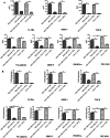
CD133+ HPCs affected the expression profiles of cytokines/chemokines/MMPs by cancer cells. HPCs were co-cultured with MDA-MB-435s-HM or MDA-MB-435s cells. The concentrations of CXCL16, IL-2Rα, IL-2Rγ, MMP-1, MMP-9, PDGFR-α, SDF-1α, TGF-β, PECAM-1, and VE-cadherin in the supernatant of MDA-MB-435s-HM cells were significantly higher than those in the supernatant of MDA-MB-435s cells (> fivefold, aj). The concentrations of MMP-9, PDGFR-α, and PECAM-1 in the supernatant of MDA-MB-435s-HM cells were significantly elevated after co-culture of MDA-MB-435s-HM cells with CD133+ HPCs cells (e, f, i). The concentrations of CXCL-16, IL-2Rα, MMP-1, MMP-9, PDGFR-α, and PECAM-1 in the supernatant of CD133+ HPCs were elevated when MDA-MB-435s-HM cells were co-cultured with CD133+ HPCs (a, b, d, f, i)
Fig. 5
Fig. 5
Validation of protein microarray results by ELISA and qRT-PCR. HPCs were co-cultured with MDA-MB-435s-HM or MDA-MB-435s cells. a The ELISA results showed trends consistent with those observed by protein microarray in the concentrations of IL-2Rα, MMP-1, MMP-9, PDGFR-α, TGF-β, PECAM-1, and VE-cadherin among the four groups. However, there were no significant differences in CXCL-16, IL-2Rγ and SDF-1α expression among the four groups. b The trends in the mRNA expression of IL-2Rα, MMP-1, MMP-9, PDGFR-α, TGF-β, PECAM-1, and VE-cadherin among the four groups were completely consistent with protein concentration results obtained by ELISA
Fig. 6
Fig. 6
Effect of the inhibition of MMP-9, PDGFR-α, and PECAM-1 on the behaviors of MDA-MB-435s-HM cells. HPCs were co-cultured with MDA-MB-435s-HM cells. a Representative images of MDA-MB-435s-HM cell migration. TIMP-1, PDGFR tyrosine kinase inhibitor III, and WM59 were added to the culture medium in the experiment group, and normal saline was added to the control group. A significantly decreased number of migrated cells could be seen in the experimental group. b Statistical analysis of cell migration. c Inhibition of MMP-9, PDGFR-α, and PECAM-1 suppresses lung metastasis of MDA-MB-435s-HM cells
Fig. 7
Fig. 7
Inhibition of MMP-9, PDGFR-α, and PECAM-1 decreases the amount of VEGFR1+CD133+ HPCs in the lung. The peak of the average ratio of VEGFR1+ HPCs was postponed and synchronous to the appearance of detectable lung metastasis. Transfection of miR-140-3p enhanced the antitumor effect of sorafenib on MHCC97-H cells’ intrahepatic growth
Fig. 8
Fig. 8
HPCs enhance the invasive growth of MDA-MB-435s-HM cells. HPCs were co-cultured with MDA-MB-435s-HM cells, and cells were treated with an inhibitor of MMP-9, anti-CD31 antibody, or an inhibitor of PDGFR-α. The invasive growth was assessed based on aMicroPET photographs, area of nodules in liver formed by MDA-MB-435s-HM cells or Masson staining indicating the invasive growth of cells. Quantitative results from bMicroPET photographs; c fold difference in radioactivity between liver and blood; d inhibitory rates of inhibitors on 18F-FDG intensity or e invasive growth of cells. Definitions: inhibitor of MMP-9, tissue inhibitor of metalloproteinase-1 (TIMP-1) shown to be a natural inhibitor of MMP-9; anti-CD31 antibody, anti-human CD31 antibody recognizing the D2 extracellular portion of CD31 to block its function; inhibitor for PDGFR-α, PDGFR tyrosine kinase inhibitor III shown to be an ATP-competitive inhibitor of PDGFR-α. *P < 0.05. White arrow in Figure indicates the nodules in mouse liver
Fig. 9
Fig. 9
HPCs enhances the invasive growth of MDA-MB-435s-HM cells. HPCs were co-cultured with MDA-MB-435s-HM cells, and cells were treated with an inhibitor of LY-294002, PD98059, SB431542, CP690550, or BIRB796. The invasive growth was assessed based on aMicroPET photographs, as area of nodules in liver formed by MDA-MB-435s-HM cells or Masson staining indicating the invasive growth of cells. Quantitative results from bMicroPET photographs; c fold difference in radioactivity between liver and blood; d inhibitory rates of inhibitors on 18F-FDG intensity or e invasive growth of cells. Definitions: inhibitor of MMP-9, tissue inhibitor of metalloproteinase-1 (TIMP-1) shown to be a natural inhibitor of MMP-9; anti-CD31 antibody, anti-human CD31 antibody recognizing the D2 extracellular portion of CD31 to block its function; inhibitor for PDGFR-α, PDGFR tyrosine kinase inhibitor III shown to be an ATP-competitive inhibitor of PDGFR-α. *P < 0.05. White arrow in Figure indicates the nodules in mouse liver

Similar articles

Cited by

References

    1. Abdollahi A, Li M, Ping G, Plathow C, Domhan S, Kiessling F, Lee LB, McMahon G, Gröne HJ, Lipson KE, Huber PE (2005) Inhibition of platelet-derived growth factor signaling attenuates pulmonary fibrosis. J Exp Med 201:925–935 - PMC - PubMed
    1. Ajibade AA, Wang Q, Cui J, Zou J, Xia X, Wang M, Tong Y, Hui W, Liu D, Su B, Wang HY, Wang RF (2012) TAK1 negatively regulates NF-κB and p38 MAP kinase activation in Gr-1 + CD11b + neutrophils. Immunity 36:43–54 - PMC - PubMed
    1. Andrae J, Gallini R, Betsholtz C (2008) Role of platelet-derived growth factors in physiology and medicine. Genes Dev 22:1276–1312 - PMC - PubMed
    1. Arvelo F, Sojo F, Cotte C (2016) Tumour progression and metastasis. Ecancermedicalscience 10:617 - PMC - PubMed
    1. Baik YH, An JY, Noh JH, Sohn TS, Kim S (2012) Can serum interleukin-2 receptor alpha predict lymph node metastasis in early gastric cancer? J Korean Surg Soc 82(3):143–148 - PMC - PubMed

MeSH terms

Substances