Review
. 2019 May;137(5):693-714.
doi: 10.1007/s00401-018-1930-z. Epub 2018 Nov 27.

Post-stroke inflammation-target or tool for therapy?

Affiliations
Review

Post-stroke inflammation-target or tool for therapy?

Kate Lykke Lambertsen et al. Acta Neuropathol. 2019 May.

Abstract

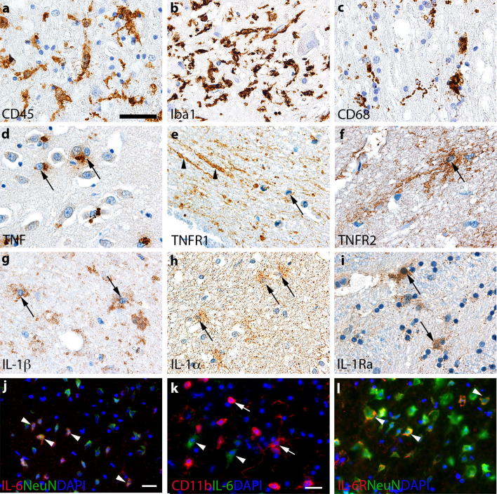
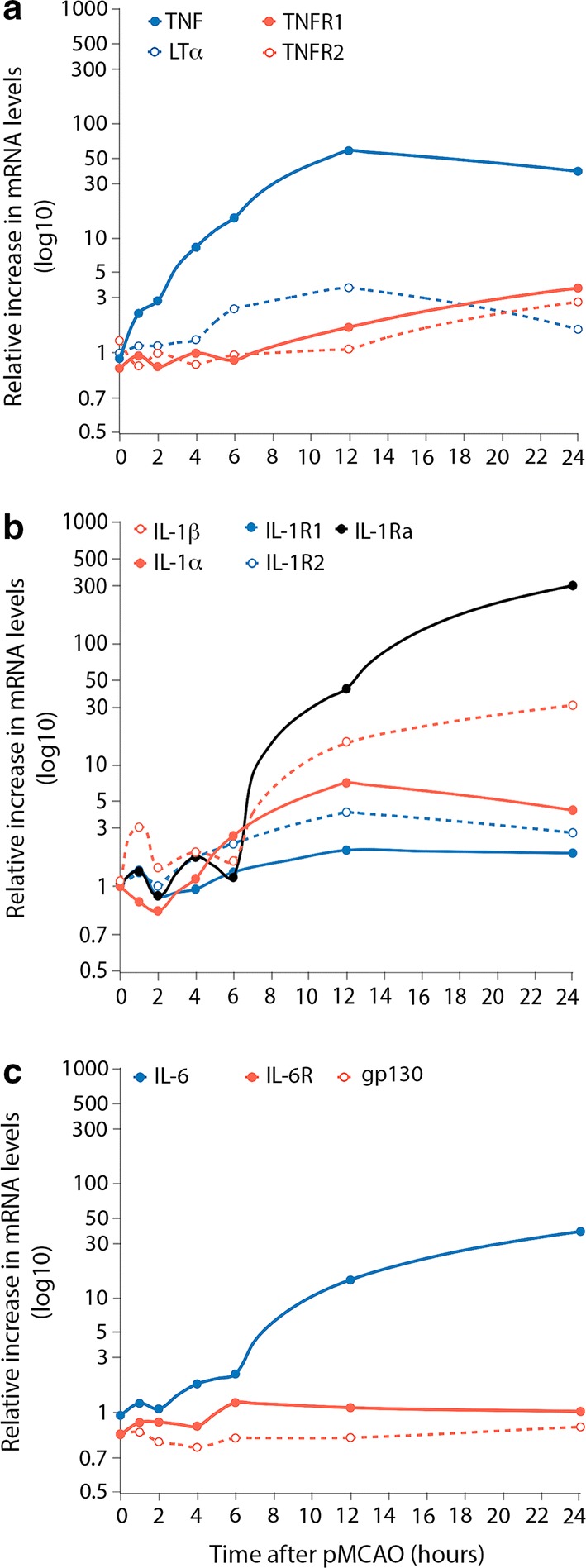
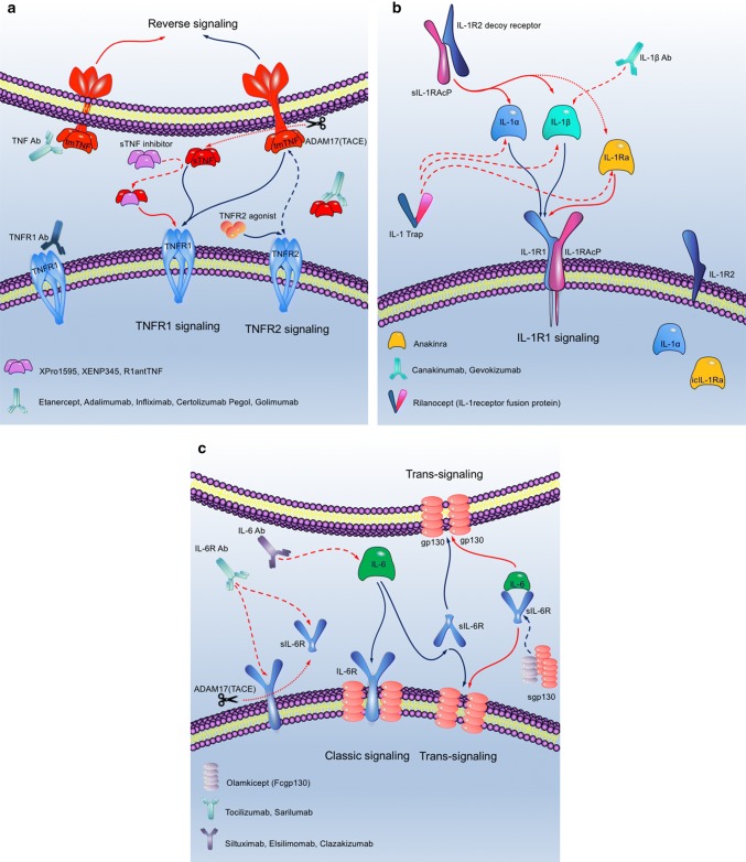
Inflammation is currently considered a prime target for the development of new stroke therapies. In the acute phase of ischemic stroke, microglia are activated and then circulating immune cells invade the peri-infarct and infarct core. Resident and infiltrating cells together orchestrate the post-stroke inflammatory response, communicating with each other and the ischemic neurons, through soluble and membrane-bound signaling molecules, including cytokines. Inflammation can be both detrimental and beneficial at particular stages after a stroke. While it can contribute to expansion of the infarct, it is also responsible for infarct resolution, and influences remodeling and repair. Several pre-clinical and clinical proof-of-concept studies have suggested the effectiveness of pharmacological interventions that target inflammation post-stroke. Experimental evidence shows that targeting certain inflammatory cytokines, such as tumor necrosis factor, interleukin (IL)-1, IL-6, and IL-10, holds promise. However, as these cytokines possess non-redundant protective and immunoregulatory functions, their neutralization or augmentation carries a risk of unwanted side effects, and clinical translation is, therefore, challenging. This review summarizes the cell biology of the post-stroke inflammatory response and discusses pharmacological interventions targeting inflammation in the acute phase after a stroke that may be used alone or in combination with recanalization therapies. Development of next-generation immune therapies should ideally aim at selectively neutralizing pathogenic immune signaling, enhancing tissue preservation, promoting neurological recovery and leaving normal function intact.

Keywords: Cytokines; Drugs; Immune therapy; Ischemia; Neuroprotection.


Figures

Fig. 1
Fig. 1
Neuroinflammation in the post-ischemic human and murine brain. ac Immunohistochemical staining of CD45+ (a), Iba1+ (b), and CD68+ (c) microglia/macrophages in human post-mortem ischemic brain tissue. di Immunohistochemical staining of TNF+ (d), TNFR1+ (e), TNFR2+ (f), IL-1β+ (g), IL-1α+ (h), and IL-1Ra+ (i) cells in human post-mortem ischemic brain tissue. (j, k) Immunofluorescence double staining showing co-localization of IL-6 to NeuN+ neurons (j), but absence of IL-6 to CD11b+ microglia/macrophages (k) in the murine brain after pMCAO. l Immunofluorescence double staining showing co-localization of IL-6R to NeuN+ neurons in the murine brain after pMCAO. Unpublished images of CD45, Iba1, CD68, TNF, TNFR1, TNFR2, and IL-1Ra stained tissue sections were acquired from human post-mortem ischemic brain tissue processed as previously described [31, 33] using already published protocols, except for IL-1β and IL-1α. Staining for IL-1β and IL-1α was performed using similar protocols and the following antibodies: Human IL-1α Ab (monoclonal mouse IgG2A, clone #4414, 1:1,200, R&D Systems) and human IL-1β Ab (monoclonal mouse IgG1, clone #2E8, 1:50, BioRad). Unpublished images of IL-6 and IL-6R co-localized cells were acquired from parallel tissue sections from mice subjected to pMCAO as described in [70]. In images ai, Toluidine blue was used as a counterstain and in jl, DAPI was used as a nuclear marker. Scale bars: a, i = 40 μm, j = 20 μm, and k, l = 20 μm. IL interleukin, IL-6R interleukin-6 receptor, TNF tumor necrosis factor, TNFR tumor necrosis factor receptor. The use of human brains was approved by the Danish Biomedical Research Ethical committee for the Region of Southern Denmark (permission number S-20080042) as stated in the original references
Fig. 2
Fig. 2
Temporal profile of cytokine and cytokine receptor upregulation in the acute phase after pMCAO. a Graphical presentation of the temporal profile of TNF, LTα, TNFR1, and TNFR2 mRNAs in the same ischemic hemispheres from mice subjected to pMCAO. b Graphical presentation of the temporal profile of IL-1β, IL-1α, IL-1Ra, IL-1R1, and IL-1R2 mRNAs after pMCAO. c Graphical presentation of the temporal profile of IL-6, IL-6R, and gp130 mRNAs after pMCAO. Data are presented as relative increases in mRNA levels compared with unmanipulated controls. TNF, TNFR1 and TNFR2 mRNA data have been obtained from [93, 94], whereas LTα mRNA data are unpublished data performed on the same experimental mice and conditions as [94]. The sequence of the LTα TaqMan probe was AGGAGGGAGTTGTTGCTCAAAGAGAAGCCA, for the LTα sense primer it was CTGCTGCTCACCTTGTTGGG, and for the LTα antisense primer it was TAGAGGCCACTGGTGGGGAT. IL-1α, IL-1β, IL-1Ra, IL-1R1, and IL-1R2 mRNA data have been obtained from [33]. IL-6, IL-6R, and gp130 mRNA data have been obtained from [70]. Note the logarithmic Y axis. gp130 glycoprotein 130, IL interleukin, IL-6R interleukin-6 receptor, LTα lymphotoxin-alpha, TNF tumor necrosis factor, TNFR tumor necrosis factor receptor
Fig. 3
Fig. 3
Schematics presenting mechanisms of actions of approved and selected experimental cytokine and cytokine receptor agonists and antagonists. ac TNF (a), IL-1 (b), and IL-6 (c) signaling via their receptors and mechanisms of actions of approved and selected novel inhibitors. Figures are modified using Protein Lounge Pathway Database (www.proteinlounge.com). Ab antibody, gp130 glycoprotein 130, icIL-1Ra intracellular interleukin-1 receptor antagonist, IL interleukin, IL-1Ra interleukin-1 receptor antagonist, IL-1R1 interleukin-1 receptor type 1, IL-1R2 interleukin-1 receptor type 2, IL-1RAcP IL-1 receptor accessory protein, sIL-1RAcP soluble IL-1 receptor accessory protein, IL-6R interleukin-6 receptor, sgp130 soluble glycoprotein 130, solIL-6R soluble interleukin-6 receptor, solTNF soluble tumor necrosis factor, tmTNF transmembrane tumor necrosis factor, TNF tumor necrosis factor, TNFR tumor necrosis factor receptor

Similar articles

Cited by

References

    1. Aderka D. The potential biological and clinical significance of the soluble tumor necrosis factor receptors. Cytokine Growth Factor Rev. 1996;7:231–240. doi: 10.1016/S1359-6101(96)00026-3. - DOI - PubMed
    1. Albers GW, Marks MP, Lansberg MG. Thrombectomy for stroke with selection by perfusion imaging. N Engl J Med. 2018;378:1849–1850. doi: 10.1056/NEJMoa1713973. - DOI - PubMed
    1. Ali C, Nicole O, Docagne F, Lesne S, MacKenzie ET, Nouvelot A, et al. Ischemia-induced interleukin-6 as a potential endogenous neuroprotective cytokine against NMDA receptor-mediated excitotoxicity in the brain. J Cereb Blood Flow Metab. 2000;20:956–966. doi: 10.1097/00004647-200006000-00008. - DOI - PubMed
    1. Allen C, Thornton P, Denes A, McColl BW, Pierozynski A, Monestier M, et al. Neutrophil cerebrovascular transmigration triggers rapid neurotoxicity through release of proteases associated with decondensed DNA. J Immunol. 2012;189:381–392. doi: 10.4049/jimmunol.1200409. - DOI - PMC - PubMed
    1. Arango-Davila CA, Vera A, Londono AC, Echeverri AF, Canas F, Cardozo CF, et al. Soluble or soluble/membrane TNF-alpha inhibitors protect the brain from focal ischemic injury in rats. Int J Neurosci. 2015;125:936–940. doi: 10.3109/00207454.2014.980906. - DOI - PubMed

Publication types