

doi: 10.1590/abd1806-4841.20187863.
Vertical plane ex vivo dermoscopy: a novel dermoscopic technique. A preliminary study
Affiliations
- PMID: 30484541
- PMCID: PMC6256214
- DOI: 10.1590/abd1806-4841.20187863
Item in Clipboard
Vertical plane ex vivo dermoscopy: a novel dermoscopic technique. A preliminary study
An Bras Dermatol.
2018 Nov/Dec.
Abstract
Dermoscopic examination allows visualization of horizontal images on the skin, showing well-defined patterns. The authors propose ex vivo dermoscopic visualization using a vertical view of skin sections. The new image clearly distinguishes the stratum corneum, epidermis, and dermis as well as the anatomical location of the pigment. Will this new approach be useful in dermoscopic diagnosis? Further studies are needed.
Conflict of interest statement
Conflict of interest: None.
Figures
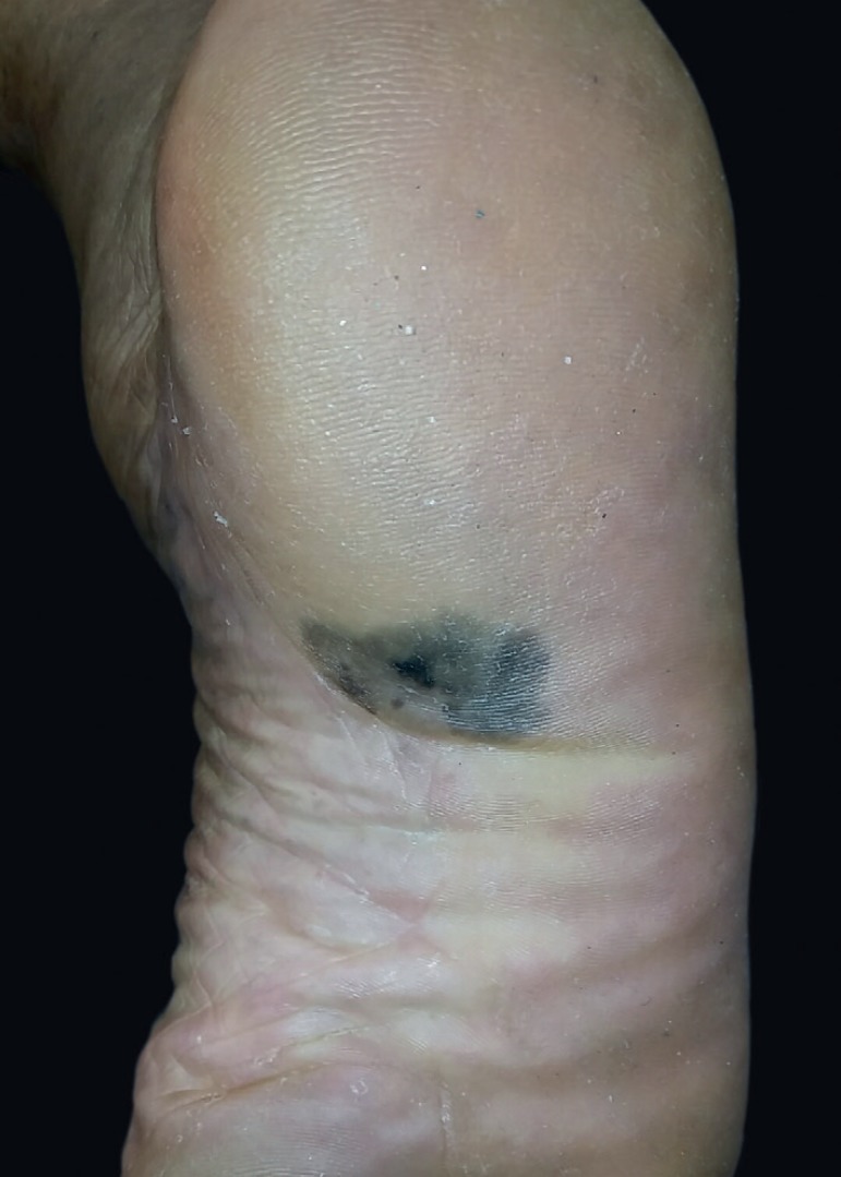
Dermatosis located on the plantar cavum characterized as an irregular macule
with multiple shades, from black to grayish, measuring 3x2cm
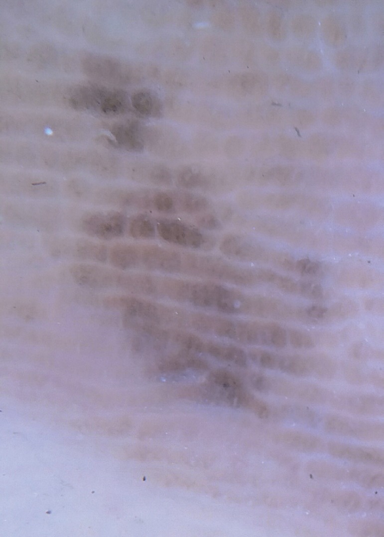
Dermoscopy showing parallel and homogeneous ridge pattern in the lesion
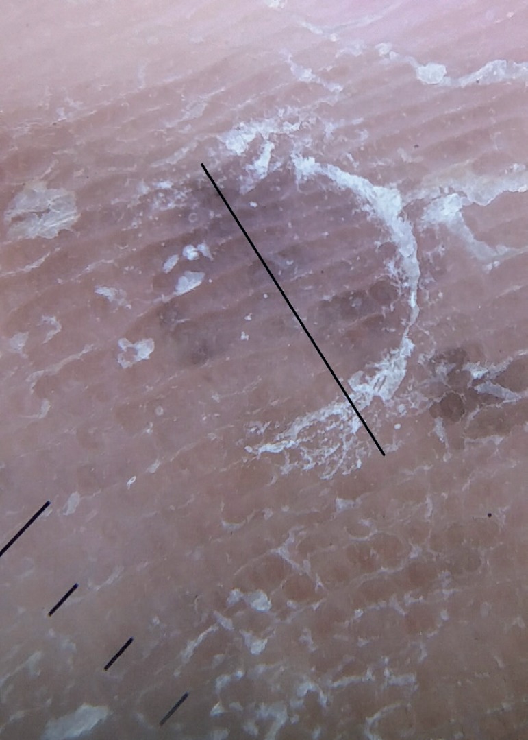
Punch biopsy (nº. 4); line represents the direction of the section in the
specimen, perpendicular to the parallel ridges
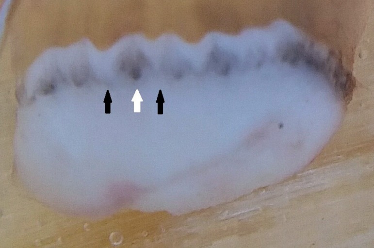
Melanin limited to the intermediate ridges and also marking the
intraepithelial duct of the sweat glands. White arrow shows the intermediate
ridge. The black arrow showing the limiting ridges
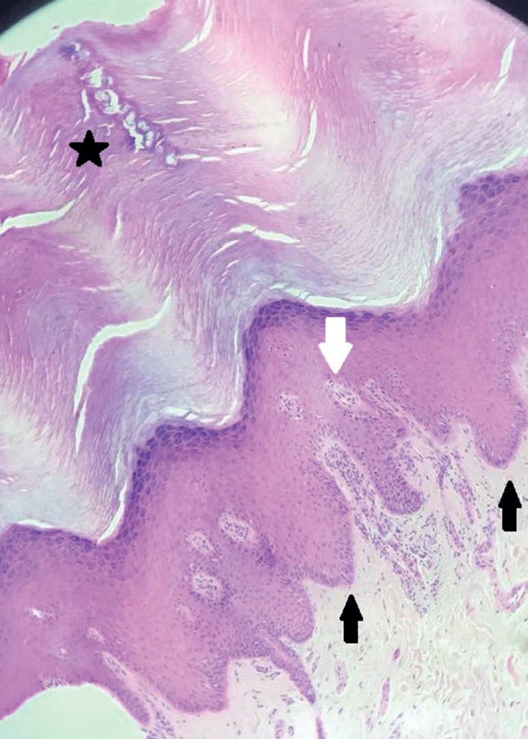
Histologic section of plantar skin shows hyperparakeratosis with melanocytic
atypia in the basal layer of epidermal ridges with pleomorphism and oriented
upwards, characterizing malignant melanocytic lesion/acral melanoma. White
arrow shows the intermediate ridge. Black arrow shows the limiting ridges.
The star represents the glandular duct
References
-
- Kittler H, Pehamberger H, Wolff K, Binder M. Diagnostic accuracy of dermoscopy. Lancet Oncol. 2002;3:159–165. - PubMed
-
- Braun RP, Thomas L, Dusza SW, Gaide O, Menzies S, Dalle S, et al. Dermoscopy of acral melanoma: a multicenter study on behalf of the International Society of Dermoscopy. Dermatology. 2013;227:373–380. - PubMed
-
- Scope A, Busam KJ, Malvehy J, Puig S, McClain SA, Braun RP, et al. Ex vivo dermoscopy of melanocytic tumors: time for dermatopathologists to learn dermoscopy. Arch Dermatol. 2007;143:1548–1552. - PubMed
-
- Braun RP, Kaya G, Masouyé I, Krischer J, Saurat JH. Histopathologic correlation in dermoscopy: a micropunch technique. Arch Dermatol. 2003;139:349–351. - PubMed
-
- Miyazaki A, Saida T, Koga H, Oguchi S, Suzuki T, Tsuchida T. Anatomical and histopathological correlates of the dermoscopic patterns seen in melanocytic nevi on the sole: a retrospective study. J Am Acad Dermatol. 2005;53:230–236. - PubMed
MeSH terms
LinkOut - more resources
Full Text Sources
Medical
