. 2019 Jan 1;155(1):58-65.
doi: 10.1001/jamadermatol.2018.4378.

Expert-Level Diagnosis of Nonpigmented Skin Cancer by Combined Convolutional Neural Networks

Affiliations

Expert-Level Diagnosis of Nonpigmented Skin Cancer by Combined Convolutional Neural Networks

Philipp Tschandl et al. JAMA Dermatol. .

Abstract

Importance: Convolutional neural networks (CNNs) achieve expert-level accuracy in the diagnosis of pigmented melanocytic lesions. However, the most common types of skin cancer are nonpigmented and nonmelanocytic, and are more difficult to diagnose.

Objective: To compare the accuracy of a CNN-based classifier with that of physicians with different levels of experience.

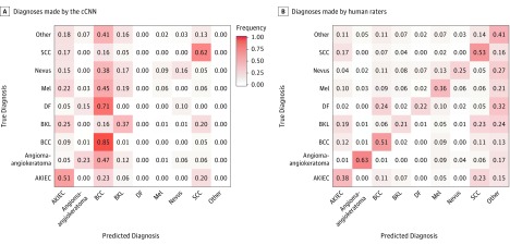
Design, setting, and participants: A CNN-based classification model was trained on 7895 dermoscopic and 5829 close-up images of lesions excised at a primary skin cancer clinic between January 1, 2008, and July 13, 2017, for a combined evaluation of both imaging methods. The combined CNN (cCNN) was tested on a set of 2072 unknown cases and compared with results from 95 human raters who were medical personnel, including 62 board-certified dermatologists, with different experience in dermoscopy.

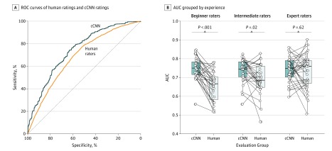
Main outcomes and measures: The proportions of correct specific diagnoses and the accuracy to differentiate between benign and malignant lesions measured as an area under the receiver operating characteristic curve served as main outcome measures.

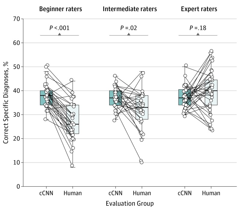
Results: Among 95 human raters (51.6% female; mean age, 43.4 years; 95% CI, 41.0-45.7 years), the participants were divided into 3 groups (according to years of experience with dermoscopy): beginner raters (<3 years), intermediate raters (3-10 years), or expert raters (>10 years). The area under the receiver operating characteristic curve of the trained cCNN was higher than human ratings (0.742; 95% CI, 0.729-0.755 vs 0.695; 95% CI, 0.676-0.713; P < .001). The specificity was fixed at the mean level of human raters (51.3%), and therefore the sensitivity of the cCNN (80.5%; 95% CI, 79.0%-82.1%) was higher than that of human raters (77.6%; 95% CI, 74.7%-80.5%). The cCNN achieved a higher percentage of correct specific diagnoses compared with human raters (37.6%; 95% CI, 36.6%-38.4% vs 33.5%; 95% CI, 31.5%-35.6%; P = .001) but not compared with experts (37.3%; 95% CI, 35.7%-38.8% vs 40.0%; 95% CI, 37.0%-43.0%; P = .18).

Conclusions and relevance: Neural networks are able to classify dermoscopic and close-up images of nonpigmented lesions as accurately as human experts in an experimental setting.


Conflict of interest statement

Conflict of Interest Disclosures: Dr Tschandl reported receiving an unrestricted grant from MetaOptima Technology Inc for conducting a 1-year postdoctoral fellowship at Simon Fraser University, Burnaby, British Columbia, Canada.

Figures

Figure 1.
Figure 1.. Comparison of Skin Cancer Detection on Digital Images Between Human Readers and a Neural Network–Based Classifier
A, Receiver operating characteristic (ROC) curves of pooled human ratings (orange) and the combined convolutional neural network (cCNN) rating (blue) show significantly higher performance by the automated classifier. B, Area under the curve (AUC) of corresponding reading sets of the cCNN and dermatologists, grouped by experience. The horizontal line in each box indicates the median (middle band), while the top and bottom borders of the box indicate the 75th and 25th percentiles, respectively.
Figure 2.
Figure 2.. Percentages of Correct Specific Diagnoses of Corresponding Reading Sets of the Combined Convolutional Neural Network (cCNN) and Dermatologists Grouped by Experience
The horizontal line in each box indicates the median (middle band), while the top and bottom borders of the box indicate the 75th and 25th percentiles, respectively.
Figure 3.
Figure 3.. Confusion Matrices of Specific Diagnoses
A, Diagnoses made by the combined convolutional neural network (cCNN). B, Diagnoses made by human raters. Values normalized to ground truth (rows). For reference, see frequency gradient scale used in panel A. AKIEC indicates actinic keratoses and intraepithelial carcinoma (also known as Bowen disease); BCC, basal cell carcinoma (all subtypes); BKL, benign keratosis-like lesions (including solar lentigo, seborrheic keratosis and lichen planus–like keratosis); DF, dermatofibroma; Mel, melanoma; and SCC, invasive squamous cell carcinoma.
Figure 4.
Figure 4.. Example Images
A, A clear cell acanthoma (CCA) correctly diagnosed by all human raters, but interpreted as a benign keratosis-like lesion by the combined convolutional neural network (cCNN). Since the class CCA was not present in the training data set it is impossible for the fixed classifier to ever make that diagnosis. B, An actinic keratosis and intraepithelial carcinoma (also known as Bowen disease) correctly specified by both the cCNN and all human raters.

Comment in

  • MUW researcher of the month.
    Tschandl P. Tschandl P. Wien Klin Wochenschr. 2019 Nov;131(21-22):582-583. doi: 10.1007/s00508-019-01580-1. Wien Klin Wochenschr. 2019. PMID: 31713738 No abstract available.

Similar articles

Cited by

References

    1. Kittler H, Marghoob AA, Argenziano G, et al. . Standardization of terminology in dermoscopy/dermatoscopy: results of the third consensus conference of the International Society of Dermoscopy. J Am Acad Dermatol. 2016;74(6):1093-1106. doi:10.1016/j.jaad.2015.12.038 - DOI - PMC - PubMed
    1. Argenziano G, Cerroni L, Zalaudek I, et al. . Accuracy in melanoma detection: a 10-year multicenter survey. J Am Acad Dermatol. 2012;67(1):54-59. doi:10.1016/j.jaad.2011.07.019 - DOI - PubMed
    1. Rosendahl C, Cameron A, Bulinska A, Williamson R, Kittler H. Dermatoscopy of a minute melanoma. Australas J Dermatol. 2011;52(1):76-78. doi:10.1111/j.1440-0960.2010.00725.x - DOI - PubMed
    1. Rosendahl C, Tschandl P, Cameron A, Kittler H. Diagnostic accuracy of dermatoscopy for melanocytic and nonmelanocytic pigmented lesions. J Am Acad Dermatol. 2011;64(6):1068-1073. doi:10.1016/j.jaad.2010.03.039 - DOI - PubMed
    1. Sinz C, Tschandl P, Rosendahl C, et al. . Accuracy of dermatoscopy for the diagnosis of nonpigmented cancers of the skin. J Am Acad Dermatol. 2017;77(6):1100-1109. doi:10.1016/j.jaad.2017.07.022 - DOI - PubMed