. 2019 Mar;67(3):498-511.
doi: 10.1002/glia.23558. Epub 2018 Nov 28.

Lipid transporter Spns2 promotes microglia pro-inflammatory activation in response to amyloid-beta peptide

Affiliations

Lipid transporter Spns2 promotes microglia pro-inflammatory activation in response to amyloid-beta peptide

Liansheng Zhong et al. Glia. 2019 Mar.

Abstract

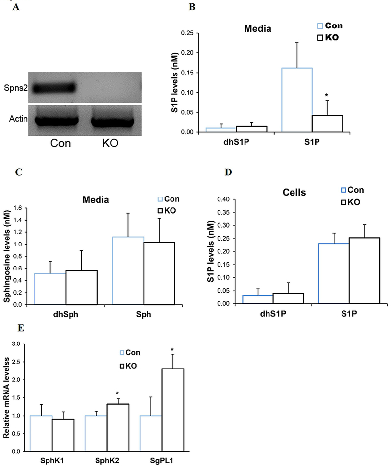
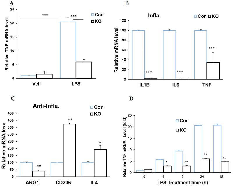
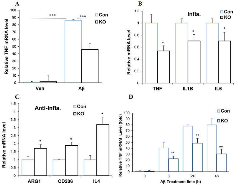
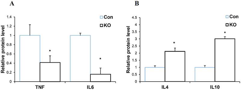
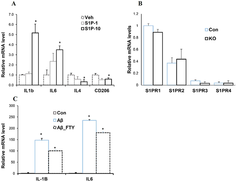
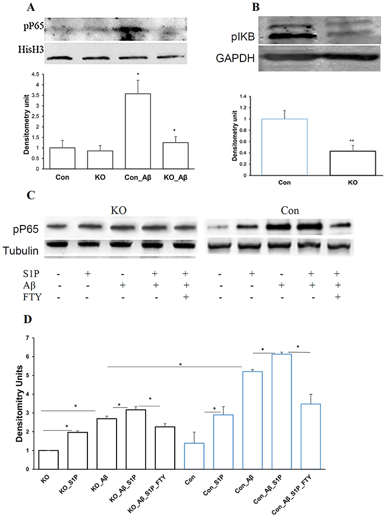
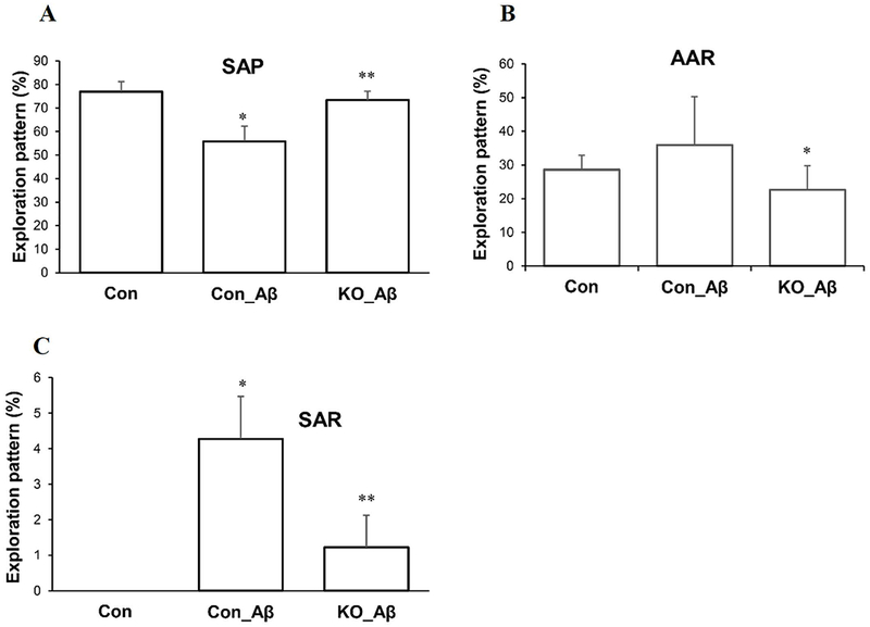
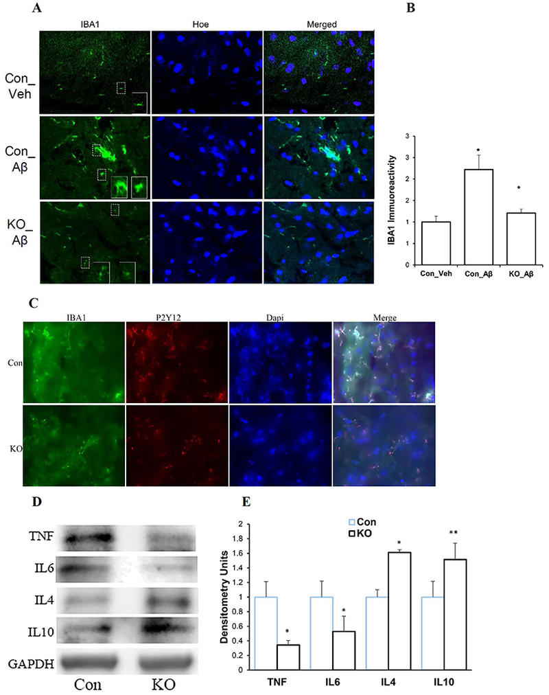
Accumulating evidence indicates that neuroinflammation contributes to the pathogenesis and exacerbation of neurodegenerative disorders, such as Alzheimer's disease (AD). Sphingosine-1-phosphate (S1P) is a pleiotropic bioactive lipid that regulates many pathophysiological processes including inflammation. We present evidence here that the spinster homolog 2 (Spns2), a S1P transporter, promotes microglia pro-inflammatory activation in vitro and in vivo. Spns2 knockout (Spns2KO) in primary cultured microglia resulted in significantly reduced levels of pro-inflammatory cytokines induced by lipopolysaccharide (LPS) and amyloid-beta peptide 1-42 oligomers (Aβ42) when compared with littermate controls. Fingolimod (FTY720), a S1P receptor 1 (S1PR1) functional antagonist and FDA approved drug for relapsing-remitting multiple sclerosis, partially blunted Aβ42-induced pro-inflammatory cytokine generation, suggesting that Spns2 promotes microglia pro-inflammatory activation through S1P-signaling. Spns2KO significantly reduced Aβ42-induced nuclear factor kappa B (NFκB) activity. S1P increased, while FTY720 dampened, Aβ42-induced NFκB activity, suggesting that Spns2 activates microglia inflammation through, at least partially, NFκB pathway. Spns2KO mouse brains showed significantly reduced Aβ42-induced microglia activation/accumulation and reduced levels of pro-inflammatory cytokines when compared with age-matched controls. More interestingly, Spns2KO ameliorated Aβ42-induced working memory deficit detected by Y-Maze. In summary, these results suggest that Spns2 promotes pro-inflammatory polarization of microglia and may play a crucial role in AD pathogenesis.

Keywords: Alzheimer's disease; Spns2; microglia; neuroinflammation; sphingosine-1-phosphate.


Conflict of interest statement

Conflict of interest: The authors declare no conflict of interest.

Figures

Figure 1.
Figure 1.
Characterization of Spns2KO microglia. A. Spns2 was expressed in littermate control primary cultured microglia (Con) while not detectable in Spns2 Knockout (Spns2KO) microglia. B. S1P was significantly reduced in the culture media of Spns2KO microglia compared to littermate Con microglia when using media with delipidated FBS, while the level of dhS1P did not change. N=3. *, p<0.05. C. The levels of Sphingosine (Sph) and dhSph were not significantly altered in Spns2KO microglia culture media. P>0.05. N=3 D. Intracellular levels of dhS1P and S1P were not significantly altered in Spns2KO microglia when compared to Con. P>0.05. N=2. E. Spns2KO differentially affected expression of S1P metabilic enzymes SphK1, SphK2, and SgPL1. *, p<0.05. N=3.
Figure 2.
Figure 2.
Spns2KO reduced pro-inflammatory polarization of microglia induced by LPS. Microglia were treated with LPS for 24 hours. A. LPS treatment significantly increased mRNA expression of TNFα when compared to vehicle (Veh). Spns2KO (KO) significantly reduced LPS-induced TNFα mRNA expression when compared to littermate control (Con). ***, p<0.001. N=3. B. Spns2KO significantly reduced expression of LPS-induced mRNA levels of TNFα, IL1b and IL6, when compared to littermate Con microglia. ***, p<0.001. N=3. C. Spns2KO significantly increased expression of LPS-induced mRNA levels of CD206 and IL4, but not ARG1, when compared to littermate Con microglia. *, p<0.05. **, p<0.01. N=3. B. Time response curve for LPS treatment.
Figure 3.
Figure 3.
Spns2KO reduced pro-inflammatory polarization and increased anti-inflammatory polarization induced by Aβ42 Oligomer (Aβ42). Microglia were treated with Aβ42 for 24 hours. A. Aβ42 treatment significantly increased mRNA expression of TNFα when compared to vehicle (Veh). Spns2KO (KO) significantly reduced Aβ42-induced TNFα mRNA expression. ***, p<0.001. N=3. B. Spns2KO significantly reduced expression of Aβ42-induced mRNA levels of TNFα, IL1b and IL6, when compared to littermate Con microglia. *, p<0.05. N=3. C. Spns2KO significantly increased expression of Aβ42-induced mRNA levels of ARG1, CD206, and IL4, when compared to littermate Con microglia. *, p<0.05. N=3.
Figure 4.
Figure 4.
Spns2KO reduced secreted pro-inflammatory and increased anti-inflammatory protein levels from microglia induced by Aβ42. A. Secreted protein levels of TNFα and IL6 were reduced in Spns2KO microglia detected by ELISA. p<0.05. N=3. B. Secreted protein levels of IL4 and IL10 were increased in Spns2KO microglia detected by ELISA. p<0.05. N=3.
Figure 5.
Figure 5.
Spns2 enhanced microglia pro-inflammatory polarization through S1P signaling. A. S1P augmented pro-inflammatory cytokine production. 1nM (S1P-1) and 10nM (S1P-10) S1P were supplemented into microglia culture. Samples were collected 1 hour later for qPCR. p<0.05. N=3. B. S1PR mRNA levels in untreated microglia. N=6-8. C. Control microglia were treated with 1μM Aβ42 oligomer for 1 hour with or without FTY-720. mRNA was extracted and IL-1β and IL6 assayed by qPCR. *, p<0.05. N=3.
Figure 6.
Figure 6.
Spns2 enhanced microglia pro-inflammatory polarization partly through NFκB signaling. A. Spns2KO reduced Aβ42-induced nuclear pP65. *, p<0.05. N=3. The bands were quantified by densitometry using ImageJ and normalized to Histone H3 (HisH3). B. Spns2KO significantly reduced Aβ42-induced pIκB level. The bands were quantified using ImageJ and normalized to GAPDH. **, p<0.01. N=3. C and D, S1P coordinated with Aβ42 to induce NFκB activity determined by Western blot (C) and densitometry analysis (D). *, p<0.05. N=3.
Figure 7.
Figure 7.
Spns2KO ameliorated Aβ42-induced cognition in vivo. Four μl of 5nM Aβ42 oligomers were injected stereotactically into the subgranular zone of 6 to 8-month-old male mouse brains. Six weeks later, the animals were subjected to Y-maze to test spatial working memory. Spns2KO ameliorated the memory decline caused by Aβ42 shown by: A, improved Spontaneous alternation performance (SAP), B, reduced alternative arm return (AAR), and C, reduced same arm return (SAR). *, p<0.05; **, p<0.01. N=4. SAP is defined as the percentage of triads that an animal goes into three different arms of the Y-maze in a triad entry; AAR as the percentage of an animal going into alternative arms in a triad entry, and SAR as the percentage of an animal returning to the same arm in any consecutive entries in a triad entry.
Figure 8.
Figure 8.
Spns2KO reduced Aβ42-induced inflammation in vivo. Four μl of 5nM Aβ42 oligomers were injected as in Figure 7. Brain samples were collected for immunofluorescence and Western Blot analyses. A. Representative images of microglia labeling of microglia in control (Con) and Spns2KO (KO) brains injected with Aβ42 and Vehicle (Veh) for IBA1 (microglia), NeuN (neuron), and Hoechst (Hoe, nuclei). B. Quantification of immunoreactivity of IBA1 in A. *, p<0.05. N=4. C. Co-labeling of microglia with IBA1 and P2Y12 in brain sections injected with Aβ42. D. Western blot to detect TNFα, IL6, IL4, and IL10 protein levels in brains injected with Aβ42. E. Densitometry quantification of the bands in D. *, p<0.05; **, p<0.01. N=3-5.

References

    1. Abduljaleel Z, Al-Allaf FA, Khan W, Athar M, Shahzad N, Taher MM, Elrobh M, Alanazi MS, El-Huneidi W. 2014. Evidence of trem2 variant associated with triple risk of Alzheimer’s disease. PLoS One 9:e92648. - PMC - PubMed
    1. Ahlers KE, Karacay B, Fuller L, Bonthius DJ, Dailey ME. 2015. Transient activation of microglia following acute alcohol exposure in developing mouse neocortex is primarily driven by BAX-dependent neurodegeneration. Glia 63:1694–713. - PMC - PubMed
    1. Alvarez SE, Harikumar KB, Hait NC, Allegood J, Strub GM, Kim EY, Maceyka M, Jiang H, Luo C, Kordula T and others. 2010. Sphingosine-1-phosphate is a missing cofactor for the E3 ubiquitin ligase TRAF2. Nature 465:1084–8. - PMC - PubMed
    1. Asai H, Ikezu S, Tsunoda S, Medalla M, Luebke J, Haydar T, Wolozin B, Butovsky O, Kugler S, Ikezu T. 2015. Depletion of microglia and inhibition of exosome synthesis halt tau propagation. Nat Neurosci 18:1584–93. - PMC - PubMed
    1. Aytan N, Choi JK, Carreras I, Brinkmann V, Kowall NW, Jenkins BG, Dedeoglu A. 2016. Fingolimod modulates multiple neuroinflammatory markers in a mouse model of Alzheimer’s disease. Sci Rep 6:24939. - PMC - PubMed

Publication types

MeSH terms