. 2019 Feb 1;30(3):400-410.
doi: 10.1091/mbc.E18-05-0297. Epub 2018 Nov 28.

Multilayered regulation of TORC1-body formation in budding yeast

Affiliations

Multilayered regulation of TORC1-body formation in budding yeast

Arron Sullivan et al. Mol Biol Cell. .

Abstract

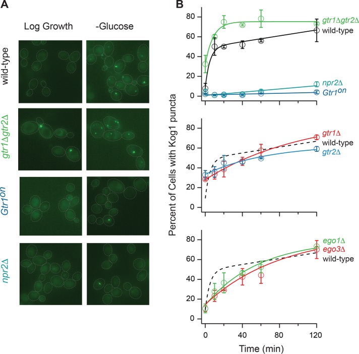
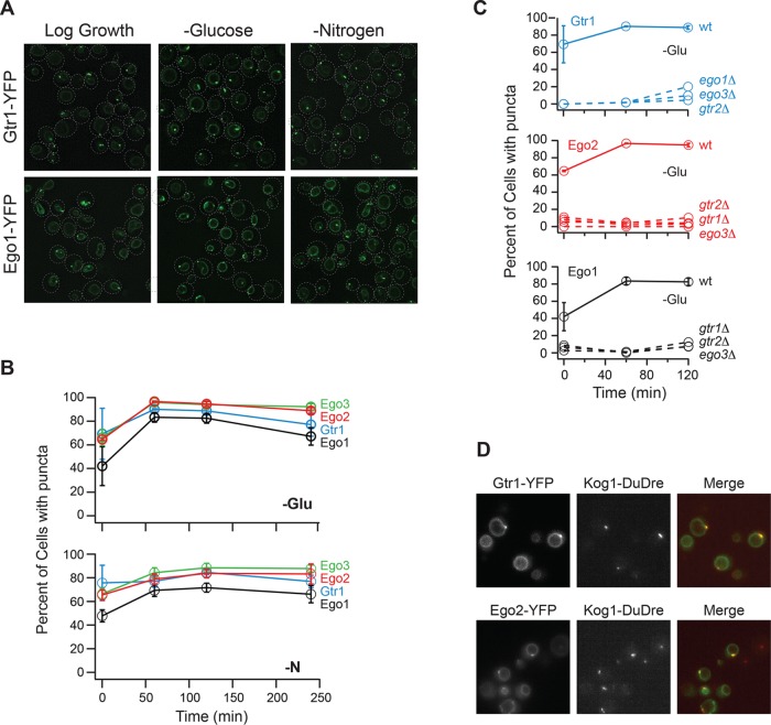
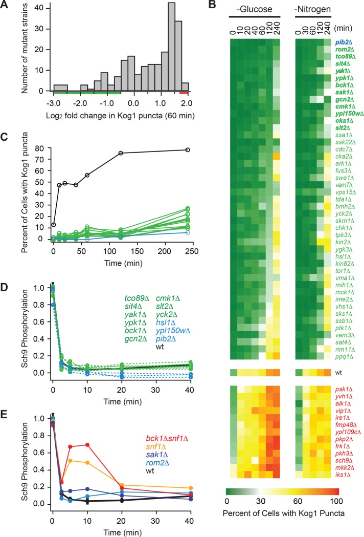
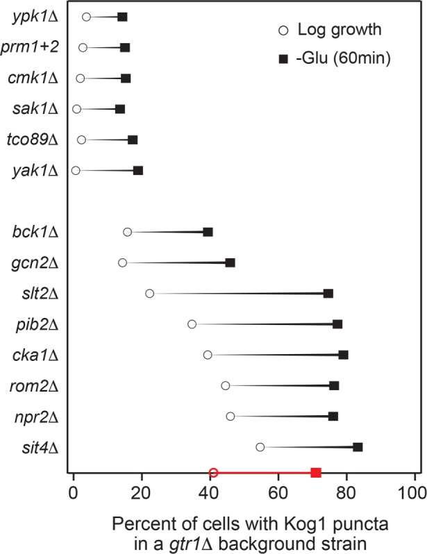
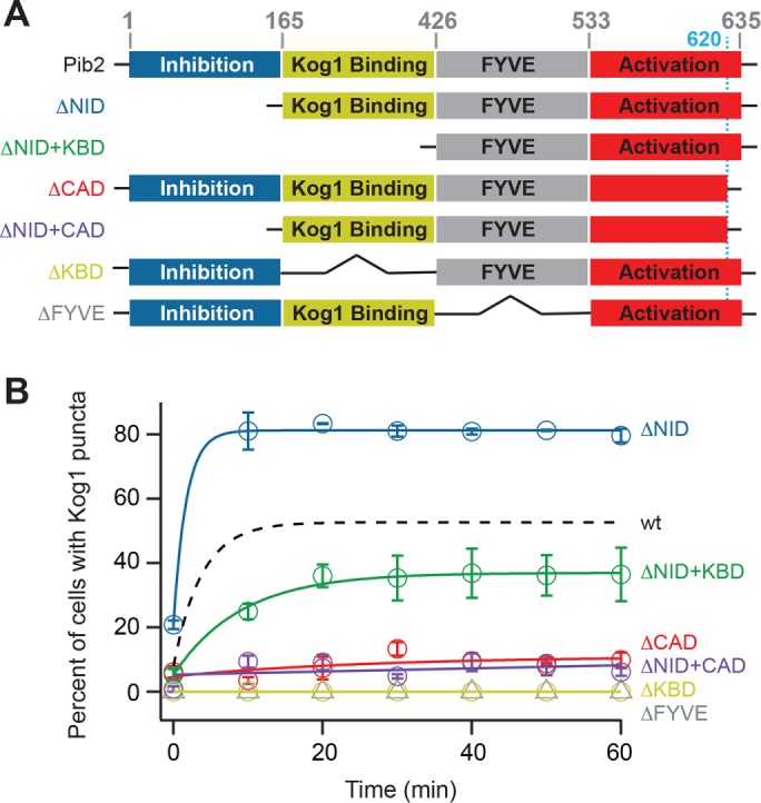
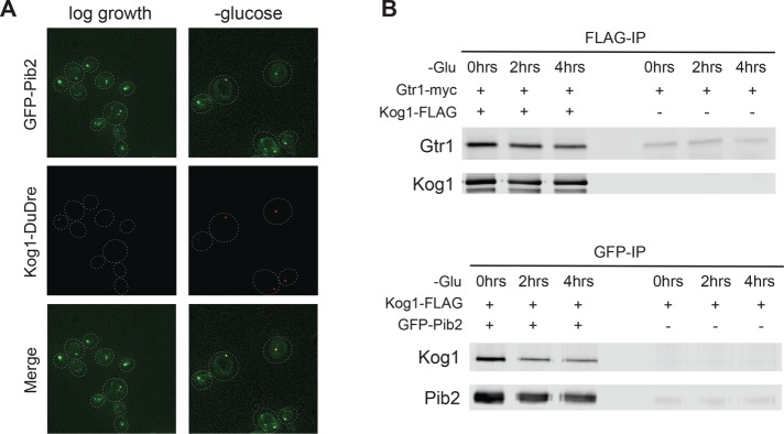
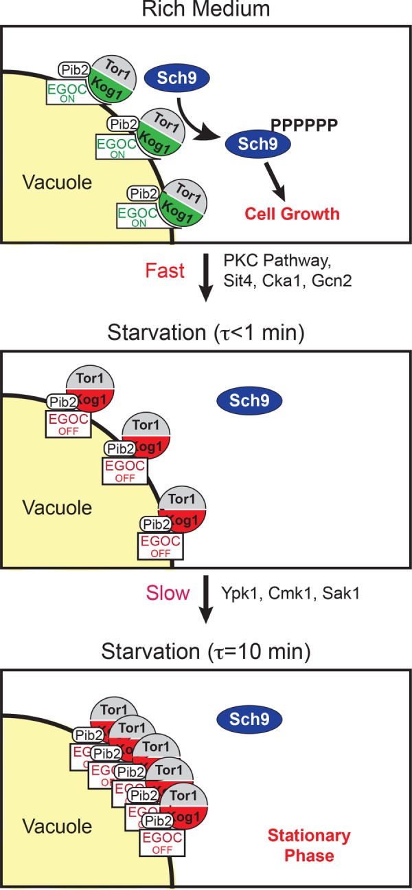
The target of rapamycin kinase complex 1 (TORC1) regulates cell growth and metabolism in eukaryotes. In Saccharomyces cerevisiae, TORC1 activity is known to be controlled by the conserved GTPases, Gtr1/2, and movement into and out of an inactive agglomerate/body. However, it is unclear whether/how these regulatory steps are coupled. Here we show that active Gtr1/2 is a potent inhibitor of TORC1-body formation, but cells missing Gtr1/2 still form TORC1-bodies in a glucose/nitrogen starvation-dependent manner. We also identify 13 new activators of TORC1-body formation and show that seven of these proteins regulate the Gtr1/2-dependent repression of TORC1-body formation, while the remaining proteins drive the subsequent steps in TORC1 agglomeration. Finally, we show that the conserved phosphatidylinositol-3-phosphate (PI(3)P) binding protein, Pib2, forms a complex with TORC1 and overrides the Gtr1/2-dependent repression of TORC1-body formation during starvation. These data provide a unified, systems-level model of TORC1 regulation in yeast.


Figures

FIGURE 1:
FIGURE 1:
EGOC regulates TORC1-body formation. (A) Kog1-YFP localization before (log growth) and 60 min after glucose starvation in the wild-type strain, and strains missing Gtr1/2 (gtr1Δgtr2Δ), Npr2 (npr2Δ), or carrying a constitutively active GTR1Q65L allele (Gtr1on). The dashed lines show the position of each cell in the bright-field image. (B) Time-course data showing the fraction of cells containing Kog1-YFP puncta in the wild-type strain and strains missing Npr2, Gtr1, Gtr2, Gtr1/2, Ego1, Ego3, or carrying a constitutively active Gtr1 allele (Gtr1on), during glucose starvation (as labeled). Each time point shows the average and SD from experiments carried out on three different days, with 75–300 cells per time point per replicate. The solid lines show the best fit to a single exponential for the gtr1Δgtr2Δ, ego1Δ, ego3Δ, gtr1Δ, and gtr2Δ strains, a double exponential for the wild-type strain, and a straight line for the npr2Δ and Gtr1on strains. The broken lines in the bottom panels show the best fit to the wild-type data (from the top panel) for comparison.
FIGURE 2:
FIGURE 2:
EGOC forms puncta in log growth and starvation conditions. (A) Gtr1-YFP and Ego1-YFP localization before (log growth), and 60 min after, glucose and nitrogen starvation. The dashed lines show the position of each cell in the bright-field image. (B) Time-course data showing the fraction of cells containing Ego1-YFP, Ego2-YFP, Ego3-YFP, and Gtr1-YFP puncta during glucose and nitrogen starvation (top and bottom panels, respectively). (C) Time-course data showing the fraction of cells containing Gtr1-YFP, Ego1-YFP, and Ego2-YFP puncta in strains missing Gtr1, Gtr2, Ego1, or Ego3 (as labeled). For B and C, each time point shows the average and SD from experiments carried out on two different days, with 100–300 cells per time point per replicate. (D) Localization of Gtr1-YFP and Kog1-DuDre (top panel) and Ego2-YFP and Kog1-DuDre (bottom panel) after 60 min of glucose starvation.
FIGURE 3:
FIGURE 3:
Screen for regulators of TORC1-body formation. (A) Histogram summarizing the influence that deleting 209 different genes (including all 139 nonessential kinases and phosphatases in yeast) has on Kog1-YFP foci formation. The score for each strain is based on the percentage of cells that form foci after 60 min of glucose starvation (based on data from at least 100 cells) and is normalized using wild-type data to calculate fold change. The raw data for each strain are included in Supplemental Table S1. (B) Heat map showing time-course data for the 45 strains that formed the fewest bodies in the initial screen (green/blue gene names), the 13 strains that formed the most bodies in the initial screen (red gene names), and the wild-type strain (wt), in glucose and nitrogen starvation conditions. Each colored square shows the fraction of cells with Kog1-puncta at a particular time point, based on images of at least 100 cells. (C) Time-course data for the 13 strains with the largest defects in body formation (green and blue lines; gene names shown in bold in B), and the wild-type strain (black line). (D, E) Quantification of band-shift data measuring Sch9 phosphorylation during glucose starvation in strains missing key regulators of TORC1-body formation. The data were normalized so that the level of Sch9 phosphorylation in the wild-type strain at time zero is set at 1.0. The raw bandshift data for each strain are shown in Supplemental Figure S3.
FIGURE 4:
FIGURE 4:
Cooperation between Gtr1 and key regulators of TORC1-body formation. Impact that deleting key regulators of TORC1-body formation, or mutating the prion domains in Kog1 (PrDm1+2), has on Kog1-YFP puncta formation in a gtr1Δ background. Black circles show the percentage of cells with bodies in log growth cultures, while black squares show the percentage of cells with bodies after 60 min of glucose starvation. The values shown are the average from experiments carried out on at least two different days with >100 cells per time point, per replicate. The SD is <5% for all mutants and time points except for sak1Δ (60 min), gcn2Δ (60 min),  pib2Δ (60 min), cka1Δ (0 min), and sit4Δ (0 min), which have standard deviations of 5–10%; npr2Δ (0 min),  sit4Δ (0 min), and sit4Δ (60 min), which have standard deviations of 10–15%; and slt2Δ (60 min) and npr2Δ (60 min), which have standard deviations of 15–20%. The red circle and square on the x-axis shows the data for the gtr1Δ single mutant for comparison.
FIGURE 5:
FIGURE 5:
Impact of Pib2 domains on TORC1-body formation. (A) Map of the different domains in Pib2 (as described in the text), and the structure of the Pib2 truncation mutants we constructed. The black lines in the ΔKBD and ΔFYVE mutants show that the neighboring domains are connected and do not represent polypeptide. (B) Time-course data following Kog1-YFP localization in strains carrying truncated forms of Pib2 at the Pib2 locus. Each time point shows the average and SD from experiments carried out on two different days, with 70–200 cells per time point per replicate (except t = 0, ΔNID+CAD, which had >40 cells per replicate). The solid lines show the best fit to a single exponential for the ΔNID, ΔNID+KBD, ΔCAD, and ΔNID+CAD strains and a straight line for the ΔKBD and ΔFYVE strains. The broken line shows the best fit to the wild-type data (from Figure 2) for comparison. Overexpression of Pib2 had little impact on TORC1-body formation; see Supplemental Figure S6 and Supplemental Text for details.
FIGURE 6:
FIGURE 6:
Pib2, EGOC, and TORC1 interact in log growth and starvation conditions. (A) Localization of GFP-Pib2 and Kog1-DuDre during log growth (left panels) and after 60 min of glucose starvation (right panels). The dashed lines show the position of each cell in the bright-field image. (B) Coimmunoprecipitation experiments following interactions between Gtr1 and Kog1 (top panel) and Pib2 and Kog1 (bottom panel) before (0 min) and after 2 and 4 h of glucose starvation. The right-hand side of each blot shows the data for a mock IP (IP from cells missing the epitope tag on Kog1 or Pib2) used to measure the background levels of Gtr1 and Kog1 in the precipitate.
FIGURE 7:
FIGURE 7:
Working model of TORC1 regulation. See the text for details.

References

    1. Adami A, Garcia-Alvarez B, Arias-Palomo E, Barford D, Llorca O. (2007). Structure of TOR and its complex with KOG1. Mol Cell , 509–516. - PubMed
    1. Aylett CH, Sauer E, Imseng S, Boehringer D, Hall MN, Ban N, Maier T. (2016). Architecture of human mTOR complex 1. Science , 48–52. - PubMed
    1. Barbet NC, Schneider U, Helliwell SB, Stansfield I, Tuite MF, Hall MN. (1996). TOR controls translation initiation and early G1 progression in yeast. Mol Biol Cell , 25–42. - PMC - PubMed
    1. Bar-Peled L, Chantranupong L, Cherniack AD, Chen WW, Ottina KA, Grabiner BC, Spear ED, Carter SL, Meyerson M, Sabatini DM. (2013). A tumor suppressor complex with GAP activity for the Rag GTPases that signal amino acid sufficiency to mTORC1. Science , 1100–1106. - PMC - PubMed
    1. Binda M, Peli-Gulli MP, Bonfils G, Panchaud N, Urban J, Sturgill TW, Loewith R, De Virgilio C. (2009). The Vam6 GEF controls TORC1 by activating the EGO complex. Mol Cell , 563–573. - PubMed

Publication types

MeSH terms

Substances