. 2018 Oct 12;131(4):1331-1338.
doi: 10.3171/2018.4.JNS172652. Print 2019 Oct 1.

In vivo histotripsy brain treatment

Affiliations

In vivo histotripsy brain treatment

Jonathan R Sukovich et al. J Neurosurg. .

Abstract

Objective: Histotripsy is an ultrasound-based treatment modality relying on the generation of targeted cavitation bubble clouds, which mechanically fractionate tissue. The purpose of the current study was to investigate the in vivo feasibility, including dosage requirements and safety, of generating well-confined destructive lesions within the porcine brain utilizing histotripsy technology.

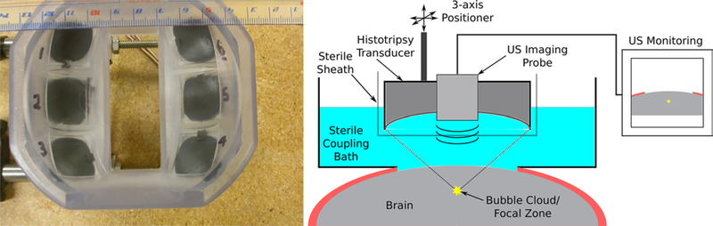
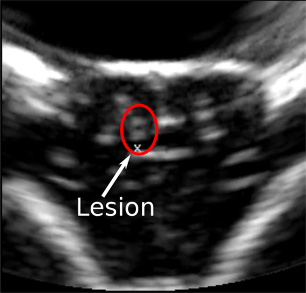
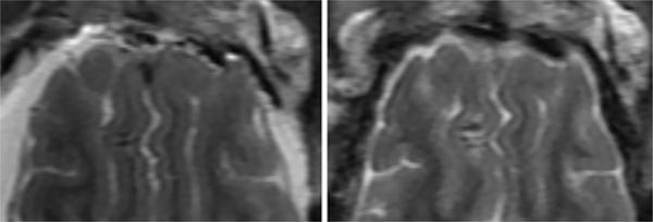
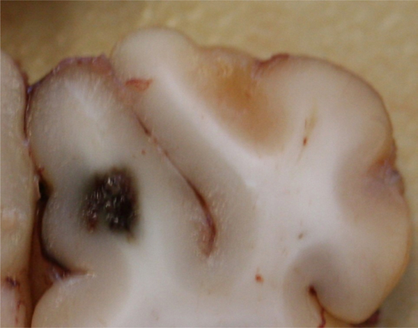
Methods: Following a craniectomy to open an acoustic window to the brain, histotripsy pulses were delivered to generate lesions in the porcine cortex. Large lesions with a major dimension of up to 1 cm were generated to demonstrate the efficacy of histotripsy lesioning in the brain. Gyrus-confined lesions were generated at different applied dosages and under ultrasound imaging guidance to ensure that they were accurately targeted and contained within individual gyri. Clinical evaluation as well as MRI and histological outcomes were assessed in the acute (≤ 6 hours) and subacute (≤ 72 hours) phases of recovery.

Results: Histotripsy was able to generate lesions with a major dimension of up to 1 cm in the cortex. Histotripsy lesions were seen to be well demarcated with sharp boundaries between treated and untreated tissues, with histological evidence of injuries extending ≤ 200 µm from their boundaries in all cases. In animals with lesions confined to the gyrus, no major hemorrhage or other complications resulting from treatment were observed. At 72 hours, MRI revealed minimal to no edema and no radiographic evidence of inflammatory changes in the perilesional area. Histological evaluation revealed the histotripsy lesions to be similar to subacute infarcts.

Conclusions: Histotripsy can be used to generate sharply defined lesions of arbitrary shapes and sizes in the swine cortex. Lesions confined to within the gyri did not lead to significant hemorrhage or edema responses at the treatment site in the acute or subacute time intervals.

Keywords: focused ultrasound; histotripsy; intracerebral hemorrhage; neurosurgery; thrombolysis.


Figures

FIG. 1.
FIG. 1.
Left: Photograph of the histotripsy transducer used to deliver treatment in these studies. The ultrasound imaging probe used during treatments for guidance and monitoring was inserted into the central slot visible in this image. Right: Schematic drawing of the setup used during treatment. US = ultrasound.
FIG. 2.
FIG. 2.
A typical ultrasound image of the brain following histotripsy treatment. The “x” below the lesion in the center of the image marks the distal end of the histotripsy focal volume and was used to target and guide lesion generation during therapy. The 2 untreated gyri to the right of the lesion, and the one to the left, appear as dark and hypo-echoic regions in ultrasonographic imaging while sulci and midline surrounding it appear bright and hyper-echoic. Following treatment, the lesion is observed as a bright, hyper-echoic region above the focal marker used to guide treatment.
FIG. 3.
FIG. 3.
Typical T2-weighted MR images of lesions generated in the brain following treatment with histotripsy. The brain shown in the image on the left was removed from the skull immediately following euthanasia, prior to decapitation. The brain shown in the image on the right was imaged in situ in the skull. Left: A coronal plane image of 3 single lesions generated by applying 10 histotripsy pulses to each target location in the left half of the brain above the ventricles. Right: An axial plane image of a block M-shaped lesion generated by steering the transducer to create the patterned lesion.
FIG. 4.
FIG. 4.
T2-weighted fast recovery fast spin echo (FRFSE) MR images of pig 4 of the subacute cohort obtained immediately following treatment (left) and at 72 hours after treatment (right). The large lesion is apparent in the cortex in the left hemisphere, the small lesion in the right hemisphere, below the large lesion.
FIG. 5.
FIG. 5.
Sagittal plane T2-cube image of the large lesion in pig 4 of the subacute cohort at 72 hours after treatment showing the presence of edema.
FIG. 6.
FIG. 6.
Photomicrographs of typical H & E–stained sections showing lesions generated using 1 (A), 10 (B), and 50 (C) histotripsy pulses. Note that the white spaces evident at the centers of the lesions are voids created as homogenized tissue fell out of the tissue during slicing.
FIG. 7.
FIG. 7.
Morphological section showing the large lesion from pig 4 of the subacute cohort. The lesion appears well confined and hemorrhagic in nature, with no evident damage or hemorrhage into the surrounding tissues.
FIG. 8.
FIG. 8.
H & E–stained sections of the large lesion generated in pig 4 of the subacute study. Left: The central region is seen to show complete tis- sue fractionation and is filled with hemorrhage and macrophages. Right: The boundary of the lesion is sharply defined, with ischemic injuries appearing to extend out 200 μm from the lesion. Adjacent tissues appear to be viable and unaffected.

References

    1. Aronow S: The use of radio-frequency power in making lesions in the brain. J Neurosurg 17:431–438, 1960 - PubMed
    1. Clement GT, White PJ, Hynynen K: Enhanced ultrasound transmission through the human skull using shear mode conversion. J Acoust Soc Am 115:1356–1364, 2004 - PubMed
    1. Cline HE, Schenck JF, Watkins RD, Hynynen K, Jolesz FA: Magnetic resonance-guided thermal surgery. Magn Reson Med 30:98–106, 1993 - PubMed
    1. Cohen-Inbar O, Xu Z, Sheehan JP: Focused ultrasound-aided immunomodulation in glioblastoma multiforme: a therapeutic concept. J Ther Ultrasound 4:2, 2016 - PMC - PubMed
    1. Coluccia D, Fandino J, Schwyzer L, O’Gorman R, Remonda L, Anon J, et al.: First noninvasive thermal ablation of a brain tumor with MR-guided focused ultrasound. J Ther Ultrasound 2:17, 2014 - PMC - PubMed

LinkOut - more resources