

Evaluation of flicker induced hyperemia in the retina and optic nerve head measured by Laser Speckle Flowgraphy
- PMID: 30485331
- PMCID: PMC6261588
- DOI: 10.1371/journal.pone.0207525
Evaluation of flicker induced hyperemia in the retina and optic nerve head measured by Laser Speckle Flowgraphy
Abstract
Purpose: The coupling between neural activity and blood flow is a physiological key principle of ocular blood flow regulation. The current study was performed to investigate whether Laser speckle flowgraphy (LSFG), a commercially available technique for measuring blood flow, is capable to assess flicker-induced haemodynamic changes in the retinal and optic nerve head (ONH) circulation.
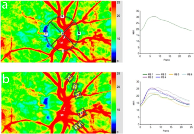
Methods: Twenty healthy subjects were included in this cross sectional study. A commercial LSFG instrument was used to measure blood flow at the ONH as well as in retinal vessels before and during stimulation with flickering light. Mean blur rate (MBR), a measure of relative blood flow velocity, was obtained for the ONH and relative flow volume (RFV) a measure of relative blood flow of the respective retinal vessels.
Results: Stimulation with flicker light increased ONH MBR by +17.5%±6.6% (p<0.01). In retinal arteries, flicker stimulation led an increase of +23.8±10.0% (p<0.05) in total RFV. For retinal veins, an increase of +23.1%±11.0 (p<0.05) in total RFV was observed during stimulation. A higher response was observed in nasal RFV compared to temporal RFV in retinal arteries (nasal: +28.9%±20.0%; temporal: +20.4%±17.6%, p<0.05) and veins (nasal: +28.3%±19.6%; temporal +17.8%±18.9%, p<0.05).
Conclusion: As shown previously with other techniques, flicker stimulation leads to an increase in retinal and optic nerve head blood flow. Our results indicate that LSFG is an appropriate method for the quantification of retinal and ONH blood flow during visual stimulation and may be used as a non-invasive, easy to use tool to assess neuro-vascular coupling in humans.
Conflict of interest statement
The authors have declared that no competing interests exist.
Figures
References
-
- Attwell D, Buchan AM, Charpak S, Lauritzen M, Macvicar BA, Newman EA. Glial and neuronal control of brain blood flow. Nature. 2010;468(7321):232–43. Epub 2010/11/12. nature09613 [pii] 10.1038/nature09613 . - DOI - PMC - PubMed
-
- Kur J, Newman EA, Chan-Ling T. Cellular and physiological mechanisms underlying blood flow regulation in the retina and choroid in health and disease. Prog Retin Eye Res. 2012;31(5):377–406. 10.1016/j.preteyeres.2012.04.004 ; PubMed Central PMCID: PMC3418965. - DOI - PMC - PubMed
-
- Newman EA. Functional hyperemia and mechanisms of neurovascular coupling in the retinal vasculature. J Cereb Blood Flow Metab. 2013. Epub 2013/08/22. 10.1038/jcbfm.2013.145 . - DOI - PMC - PubMed
-
- Riva CE, Logean E, Falsini B. Visually evoked hemodynamical response and assessment of neurovascular coupling in the optic nerve and retina. Prog Retin Eye Res. 2005;24(2):183–215. 10.1016/j.preteyeres.2004.07.002 . - DOI - PubMed
Publication types
MeSH terms
Grants and funding
LinkOut - more resources
Full Text Sources
Other Literature Sources
Research Materials
