

Microanatomical changes and biomolecular expression at the PDL-entheses during experimental tooth movement
- PMID: 30485431
- PMCID: PMC6465084
- DOI: 10.1111/jre.12625
Microanatomical changes and biomolecular expression at the PDL-entheses during experimental tooth movement
Abstract
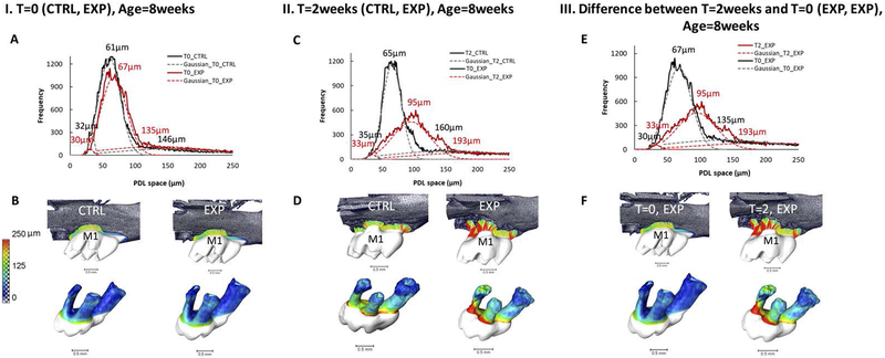
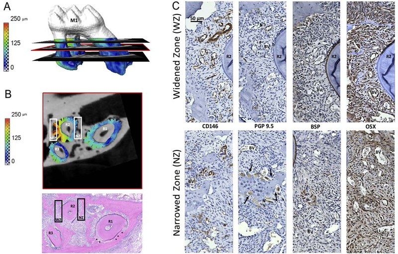
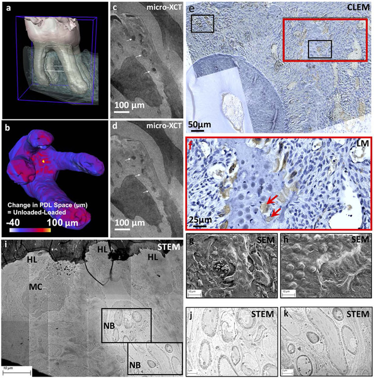
The novel aspect of this study was to contextualize the co-localization of biomolecular expression in widened and narrowed periodontal ligament (PDL)-space within a mechanically activated periodontal complex. The PDL is unique as it is the only ligament with both innervation and vascularization. Maxillary molars in 6-week-old male C57BL/6 mice (N = 5) were experimentally translated for 2 weeks using an elastic spacer. Contralateral teeth were used as controls. Mechanical testing of the periodontal complex of a mouse in situ and imaging using X-ray micro-computed tomography (micro-XCT) illustrated deformations within blood vessels (BV) of the PDL. PDL-bone and PDL-cementum entheses at the widened and narrowed PDL-spaces following experimental tooth movement (ETM) illustrated osterix (OSX), bone sialoprotein (BSP), cluster of differentiation 146 (CD146), and protein gene product 9.5 (PGP9.5), indicating active remodeling at these sites. PGP9.5 positive nerve bundles (NBs) were co-localized with multinucleated cells (MCs), Howship's resorption lacunae, and CD146 positive BVs. Association between nerves and MC was complemented by visualizing the proximity of osmium tetroxide stained NBs with the ultrastructure of MCs by performing scanning transmission electron microscopy. Spatial association of NB with BV, and NB with MC, provided insights into the plausible co-activation of NBs to initiate osteoclastic activity. Resorption of mineral occurred as an attempt to restore PDL-space of the load-bearing complex, specifically at the PDL-entheses. Mapping of anatomy-specific structural elements and their association with regenerative molecules by correlating light and electron micrographs provided insights into the use of these extracellular matrix molecules as plausible targets for pharmacological interventions related to tooth movement. Within the realm of tissue regeneration, modulation of load can reverse naturally occurring mineral formation to experimentally induced resorption, and naturally occurring mineral resorption to experimentally induced formation at the enthesial sites to permit tooth translation.
Keywords: biomolecules; enthesis; orthodontics; periodontal ligament; tooth movement.
© 2018 John Wiley & Sons A/S. Published by John Wiley & Sons Ltd.
Figures

Similar articles
-
Micro-anatomical responses in periodontal complexes of mice to calibrated orthodontic forces on the crown.Orthod Craniofac Res. 2017 Jun;20 Suppl 1:100-105. doi: 10.1111/ocr.12172. Orthod Craniofac Res. 2017. PMID: 28643923
-
Differentiating zones at periodontal ligament-bone and periodontal ligament-cementum entheses.J Periodontal Res. 2015 Dec;50(6):870-80. doi: 10.1111/jre.12281. Epub 2015 Jun 1. J Periodontal Res. 2015. PMID: 26031604 Free PMC article.
-
Effect of experimental tooth movement on nerve fibres immunoreactive to calcitonin gene-related peptide, protein gene product 9.5, and blood vessel density and distribution in rats.Eur J Orthod. 1997 Oct;19(5):517-29. doi: 10.1093/ejo/19.5.517. Eur J Orthod. 1997. PMID: 9386338
-
A Force on the Crown and Tug of War in the Periodontal Complex.J Dent Res. 2018 Mar;97(3):241-250. doi: 10.1177/0022034517744556. Epub 2018 Jan 24. J Dent Res. 2018. PMID: 29364757 Free PMC article. Review.
-
Biomechanical pathways of dentoalveolar fibrous joints in health and disease.Periodontol 2000. 2020 Feb;82(1):238-256. doi: 10.1111/prd.12306. Periodontol 2000. 2020. PMID: 31850635 Review.
Cited by
-
Advancements in composition and structural characterization of bone to inform mechanical outcomes and modelling.Curr Opin Biomed Eng. 2019 Sep;11:76-84. doi: 10.1016/j.cobme.2019.09.011. Epub 2019 Sep 28. Curr Opin Biomed Eng. 2019. PMID: 32864522 Free PMC article.
-
Periodontal ligament and alveolar bone remodeling during long orthodontic tooth movement analyzed by a novel user-independent 3D-methodology.Sci Rep. 2023 Nov 14;13(1):19919. doi: 10.1038/s41598-023-47386-0. Sci Rep. 2023. PMID: 37964111 Free PMC article.
-
Neural Regulations in Tooth Development and Tooth-Periodontium Complex Homeostasis: A Literature Review.Int J Mol Sci. 2022 Nov 16;23(22):14150. doi: 10.3390/ijms232214150. Int J Mol Sci. 2022. PMID: 36430624 Free PMC article. Review.
-
Chewing-Activated TRPV4/PIEZO1-HIF-1α-Zn Axes in a Rat Periodontal Complex.J Dent Res. 2025 Apr;104(4):398-407. doi: 10.1177/00220345241294001. Epub 2025 Jan 28. J Dent Res. 2025. PMID: 39876056 Free PMC article.
-
Alendronate partially rescues the periodontal defects in OIM mouse model of osteogenesis imperfecta.Sci Rep. 2025 Jan 2;15(1):88. doi: 10.1038/s41598-024-84756-8. Sci Rep. 2025. PMID: 39747677 Free PMC article.
References
-
- Belting CM, Schour I, Weinmann JP, Shepro MJ. Age changes in the periodontal tissues of the rat molar. J Dent Res 1953; 32(3): 332–353. - PubMed
-
- Sved A The mesial drift of teeth during growth. Am J Orthod 1955; 41(7): 539–553.
-
- Cate T, Nanci A. Ten Cate’s - Oral Histology: Development, Structure, and Function. 8th Ed. 2012. St. Louis, MO: Elsevier.
-
- Pal A, Chen L, Yang L, Yang F, Meng B, Jheon A, Ho SP. Micro-anatomical responses in the periodontal complex to calibrated orthodontic forces on the crown. Orthod Craniofac Res 2017; 20(S1): 100–105. - PubMed
MeSH terms
Substances
Grants and funding
LinkOut - more resources
Full Text Sources
Research Materials
Miscellaneous
